
Page 21 - Journal of Special Operations Medicine - Fall 2016
P. 21
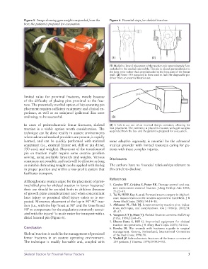
Figure 5  Image showing gym weights suspended from the   Figure 6  Essential steps for skeletal traction.
               box; the patient is prepared for evacuation.


                                                                    (1)                     (2)

                                                                  (1) Medial to lateral placement of the traction pin approximately 3cm
                                                                  cephalad to the medial epicondyle. The pin is placed perpendicular to
                                                                  the knee joint rather than perpendicular to the long axis of the femur
                                                                  itself. (2) Some 550 paracord is then used to lash the disposable pin
                                                                  driver from an external fixation set.


               limited value for proximal fractures, mostly because
               of the difficulty of placing pins proximal to the frac-
               ture. The potentially morbid option of hip-spanning pin
               placement requires sufficient equipment and clinical ex-
               perience, as well as an uninjured ipsilateral iliac crest
               and wing, to be successful.                          (3)

               In cases of peritrochanteric femur fractures, skeletal   (3) A hole is cut out of an inverted sharps container, allowing for
               traction is a viable option worth consideration. The   foot placement. The extremity is placed in traction with gym weights
               technique can be done readily in austere environments   suspended from the box and the patient is prepared for evacuation.
               where advanced medical providers are present, is rapidly
               learned, and can be quickly performed with minimal   some adaptive ingenuity, is essential for the  advanced
               equipment (i.e., external fixator set, drill or pin driver,   medical provider with limited resources caring for pa-
               550 cord, and weights). Placement of the transfemoral   tients with these complex injuries.
               pin on traction might require some creative problem
               solving, using available lanyards and weights. Various   Disclosures
               constructs are possible, and each will be effective as long
               as suitable distracting weight can be applied with the leg   The authors have no financial relationships relevant to
               in proper position and within a low-profile system that   this article to disclose.
               facilitates transport.
                                                                  References
               Although some sources argue for the placement of prox-
               imal tibial pins for skeletal traction in femur fractures,    1.  Gordon WT, Grijalva S, Potter BK. Damage control and aus-
                                                               6
               their use should be avoided both in children (because   tere environment external fixation. J Surg Orthop Adv. 2012;
                                                                     21:22–31.
               of growth plate considerations) and when concomitant   2.  Tai N, Hill P, Kay A, et al. Forward trauma surgery in Afghani-
               knee injury or proximal tibial injury exists or is sus-  stan: lessons learnt on the modern asymmetric battlefield. J R
               pected. Moreover, placement of the leg in 90°-90° trac-  Army Med Corps. 2008;154:14–18.
               tion (i.e., with the hip flexed at 90° and the knee flexed   3.  Althausen PL, Hak DJ. Lower extremity traction pins: indica-
               90° to compensate for the angulation commonly associ-  tions, technique, and complications. Am J Orthop. 2002;31:
                                                                     43–47.
                                 7
               ated with the injury)  is made easier for transport with a   4.  Vangness CT Jr, Hunt TJ. Skeletal fixation: a review. Bull Hosp
               distal femoral pin (Figure 6).                        Jt Dis. 1993;52:44–45.
                                                                  5.  Dharm-Datta  S,  Hill  G.  Improvised  equipment  for  skeletal
                                                                     traction on operations. J R Army Med Corps. 2007;154:144.
               Conclusion                                         6.  Rowley DI. War wounds with fractures: a guide to surgical
                                                                     management. Geneva, Switzerland; International Committee
               Skeletal traction is useful in the management of proximal   of the Red Cross; 1996:38.
               femur fractures in an austere operating environment.   7.  Waddell JP. Subtrochanteric fractures of the femur: a review of
               The technique is readily learnable and, coupled  with   130 patients. J Trauma. 1979;19:582–592.


               Skeletal Traction for Proximal Femur Fracture                                                     3