
Page 19 - Journal of Special Operations Medicine - Spring 2015
P. 19
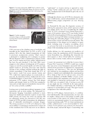
Figure 3 Coronary angiogram. (Left) Total occlusion of the “gatekeeper” to invasive testing as opposed to stress
proximal LAD at the vulnerable plaque site shown on CCTA. testing. Further studies this past year have led to the
3
(Right) Successful revascularization of the LAD shows the same conclusion that CCTA should be given the role of
large LAD territory. gatekeeper. 4
Although the effective use of CCTA for obstructive dis
ease is well known, current advancements into further
differentiating plaque composition are now showing
promise.
As illustrated by this case, the diagnostic accuracy of
defining low attenuation areas, which represents a lipid
core plaque (LCP), was key in deciphering the culprit
lesion. In 2013, European Heart Journal showcased a
study of 446 patients whose CCTA was further looked
Figure 4 Cardiac magnetic at for LCP; it was found that CCTA was a good diagnos
resonance image of subendocardial tic tool for locating LCP within coronary anatomy (i.e.,
5
scarring shown by late gadolinium identifying the vulnerable plaque). CCTA permits eval
enhancement. uation of the arterial wall and not just the lumen. The
characteristics of the plaque can be looked at in great
detail, including areas of positive remodeling, which
is a process that tends to accommodate the growth of
Discussion
plaque while minimizing luminal encroachment. 6
CAD continues to be a leading cause of morbidity and
mortality in the United States. In 50% to 65% of all Further delving into risk assessment and prediction of
patients, MI is the first clinical presentation of CAD further events, the combination of LCP detection with
in previously asymptomatic patients. Much research serum biomarkers (i.e., identifying the vulnerable pa
1
is dedicated to establishing new techniques to predict tient) appears to show clinical utility for assessing im
MI and diagnose CAD in asymptomatic patients. In the minent risk of cardiac events, such as in our patient.
past, invasive testing involving cardiac catheterization
has been the gold standard in this field. With recent A recent risk assessment tool called the Coronary Heart
progress in the technical development of CT scanners, Disease Risk Assessment (CHDRA) model was created
images can now be acquired rapidly and with very high that uses seven serum biomarkers associated with the
spatial resolution, providing physicians with detailed atherosclerotic processes of inflammation, angiogenesis,
analysis of the coronary anatomy without the risks of apoptosis, and chemotaxis. This test was developed
7
invasive procedures. It has been found that 25% of and validated in two population cohorts and has been
heart attacks result from severe stenotic lesions and shown to identify more individuals who experienced an
75% result from ruptured plaque at a moderately ste MI as being at high risk for such an event, versus the
notic site. In population studies, results from the recent National Institutes of Health (NIH)’s 10year cardio
CONFIRM Registry of more than 17,000 patients who vascular disease risk assessment tool, which is the cur
underwent CCTA analysis showed that both plaque rent standard of measuring cardiovascular risk. Using
burden and stenosis found on CCTA have prognostic the NIH tool, our patient’s risk of having an acute MI
value and can help improve risk prediction beyond clini on any single day was 1:50,100. In the same patient,
cal risk scores currently available. 2 the CHDRA identified a 5fold higher risk of MI than
expected for an individual of his age. This again leads to
Looking more in depth into traditional measures of risk recognition of the vulnerable patient.
assessment, such as stress testing, The Advanced Car
diovascular Imaging Consortium conducted a study With the CCTA plaque characterization, in combina
using their multicenter statewide registry of more than tion with biomarker risk assessment data, we were able
6000 patients and found that stress test findings did to predict a future event in this patient as well as the
not adequately predict the obstructive CAD that was exact location of the rupture site within 4 days of the
found on CCTA, nor did it demonstrate the ubiquitous event in an asymptomatic patient. In current guidelines,
ness of vulnerable nonobstructive coronary plaques. luminal narrowing under 70% by visual assessment on
The strong association of CCTA with obstructive CAD cardiac catheterization is not recommended for inter
and its ability to demonstrate nonobstructive lesions led vention. In this patient’s case, however, the characteris
8
to the conclusion that CCTA may serve as an effective tic of the plaque, and not the degree of narrowing, was
Case Report: CCTA With Biomarker Testing to Detect ACS 9