
Page 18 - Journal of Special Operations Medicine - Spring 2015
P. 18
pathways (chemotaxis, apoptosis, cell proliferation, in clopidogrel loading dose of 600mg with 75mg daily,
flammation, and angiogenesis) underlying vulnerable atorvastatin 40mg daily, and metoprolol succinate
plaque progression. The ECG revealed a preserved ejec 50mg daily. The patient was scheduled for left heart car
tion fraction at 83% with normal left ventricular (LV) diac catheterization with angioplasty and stenting the
contractility and no significant wall motion abnormali following week because of the holidays, although he did
ties. Valve morphology was also preserved. not meet national guidelines criteria. He was advised to
initiate medical therapy immediately. The patient’s wife,
CCTA was performed with a Philips 64slice scanner a nurse, was brought online remotely to review the im
(http://www.healthcare.philips.com/) with a prospective ages with the patient and the cardiologist. The patient
protocol and iterative reconstruction to minimize radia filled his prescriptions but was not going to start them
tion to less than 4 millisieverts (mSv). A total of 100mL until after the weekend. Approximately 4 days after the
of ISOVUE 370 contrast was injected in the antecubital outpatient visit, the patient began having chest discom
vein. The images were processed with Vital Images Vit fort while playing video games. The discomfort contin
rea SUREPlaque software for coronary plaque analy ued for longer than 30 minutes. He thought it was his
®
sis. The images revealed a right dominant system with fractured clavicle and did not want to seek medical help.
anatomically appropriate origins of the left and right Remembering the results of the online presentation of
coronary systems. As depicted in Figure 1, the proximal the CCTA, the patient’s wife called emergency medical
portion of the LAD showed a noncalcified plaque at the services (EMS) for further evaluation. An ECG by EMS
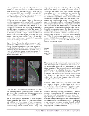
level of the first diagonal branch. personnel revealed an STelevation MI (STEMI) in the
anterior lateral leads (Figure 2).
Figure 1 Green = lumen; blue = fibrous plaque; red = lipid-
laden plaque. (Left) Zoomed-in short-axis of the LAD vessel
showing significant plaque burden with a large amount of
liquid lipid plaque (red), which was in contact with the lumen Figure 2
of the vessel, posing an imminent threat of a heart attack if ECG
it were to break through. (Right) Luminal narrowing in the showing
longitudinal view with a diameter stenosis of 54% and a acute anterior
cross-sectional area of 86%. wall MI.
The sevenprotein biomarker results were not available
until after the patient’s hospitalization because of his de
lay in getting the tests done and shipment to the West
Coast laboratory. They revealed that the patient had
an 8.07% risk of an MI within the next 5 years, which
placed him into a highrisk category. He was at a 5.34
fold higher risk of a cardiovascular event than expected
for a 48yearold man. The lipid panel revealed abnor
malities with a highdensity lipoprotein level of 31mg/dL
and triglycerides of 526mg/dL.
Emergent cardiac catheterization was performed with
the angiogram showing a total occlusion of the LAD at
the level of the vulnerable plaque. The patient under
went angioplasty and stenting of the LAD (Figure 3).
There was also a moderately calcified plaque with posi
tive remodeling in the midsegment with minimal dis The ECG during the initial STEMI showed a markedly
ease distally. The circumflex and right coronary arteries depressed ejection fraction (EF) with akinesis of the an
showed mild nonobstructive disease on CCTA. terior lateral walls extending down to the apex. Forty
eight hours post percutaneous coronary intervention,
Due to the concerning appearance of the noncalcified there was complete resolution of the abnormal territo
plaque as meeting the criteria of a vulnerable plaque, ries with normalization of LV function. At 6 weeks post
and without the information of the sevenprotein event, magnetic resonance imaging showed residual
risk assessment, the patient was prescribed aggressive subendocardial extensive anterior wall and septal scar
medical therapy, which included aspirin 325mg daily, ring (Figure 4). The EF was normal.
8 Journal of Special Operations Medicine Volume 15, Edition 1/Spring 2015