
Page 93 - Journal of Special Operations Medicine - Winter 2014
P. 93
www.idexx.com), according to manufacturer’s recom- Statistical Analysis
mendations. Peptide-specific antibody to A. phago- Local prevalence for single disease status was calculated
cytophilum, A. platys, E. canis, E. chaffeensis, or B. as the proportion of positive samples from the total of
burgdorferi present in the patient sample binds to the the samples tested. The odds ratios (ORs) for single dis-
peptide-HRP conjugate. Immune complexes that form eases among the three populations were compared by
are bound to peptide conjugates adhered to the mem- logistic regression analysis using the PROC LOGISTIC
brane on the flow matrix. Unbound antibodies and function of the Statistical Analysis System (SAS version
peptide-HRP conjugates were washed and substrate 9.2; SAS Institute Inc., Cary, NC, USA; http://www.sas
reagents were applied. Color development in the area .com/).
of the deposition species-specific peptides and immune
complexes indicated a positive result.
Results
DNA Extraction and Real-Time Of the 218 serum samples tested, 62 (28.4%) had an-
Polymerase Chain Reaction Amplification tibodies only to E. canis and 15 (6.9%) had antibod-
DNA was extracted from canine whole blood using a ies only to A. platys. However, 101 (46.3%) samples
commercially available kit (High Pure PCR Template had antibodies to both E. canis and A. platys. There-
Preparation Kit, Roche Applied Science, Madison, WI, fore, the total number of E. canis positive samples was
USA; lifescience.roche.com) according to the manufac- 163 (74.4%) and the total number of A. platys positive
turer’s instructions. Genomic DNA was stored at −20°C samples was 116 (53.2%). Of the 218 DNA samples,
until testing. 46 (21.1%) were positive for E. canis DNA, 19 (8.7%)
were positive for A. platys DNA and 16 (7.3%) were
Real-time polymerase chain reaction (PCR) hybridiza- positive for both E. canis and A. platys DNA. There-
tion probe assays were used that detect the disulfide fore, 62 samples (28.4%) were positive for the presence
oxidoreductase gene (GenBank AF403710) of E. canis of E. canis DNA and 35 samples (16.1%) were positive
and the p44 gene of A. platys. These two assays were for the presence of A. platys DNA. Of the 163 dogs se-
used based on the high seroprevalence of exposure to ropositive for E. canis, 60 (36.8%) were also positive by
these agents in the sample population. The real-time PCR analysis. Two samples were PCR positive for E. ca-
PCR assays were performed with the LightCycler 480 nis but seronegative. Of the 116 dogs seropositive for A.
instrument (Roche Applied Science, Madison, WI, USA; platys, 26 (22.4%) were also positive by PCR analysis.
lifescience.roche.com). PCR was carried out in a total re- Nine samples were PCR positive but seronegative. Of
action volume of 20μL containing LightCycler 480 Ge- the 101 samples that were seropositive for both E. canis
notyping Master mix (Roche Applied Science, Madison, and A. platys, 10 (10.0%) were also PCR positive for
WI, USA; lifescience.roche.com), assay specific primers both organisms. All 218 serum samples were seronega-
and probes, and 5μL of template DNA. Cycling param- tive for E. ewingii, E. chaffeensis, and B. burgdorferi.
eters for the E. canis PCR consisted of a denaturation Two samples were seropositive for A. phagocytophilum,
cycle of 95°C for 10 minutes, followed by a 55- cycle and both of these were PCR negative. These data are
amplification profile (95°C for 20 seconds, 60°C for outlined in Table 1. Exposure to tick-borne pathogens
30 seconds with a single data acquisition, 72°C for 20 was highest in shelter animals and military working
seconds), a melting curve profile (95°C for 1 minute, dogs: More than 90% of the samples were seropositive
45°C for 1 minute, and 80°C continuous with a ramp or PCR positive for one or more organisms as compared
rate of 0.14°C per second and four data acquisitions per to 51% in client-owned animals.
°C). The A. platys PCR cycling parameters were a de-
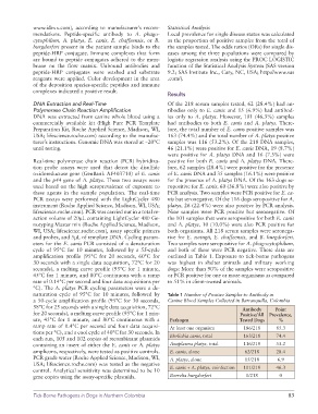
naturation cycle of 95°C for 10 minutes, followed by Table 1 Number of Positive Samples to Antibody in
a 50-cycle amplification profile (95°C for 30 seconds, Canine Blood Samples Collected in Barranquilla, Colombia
58°C for 25 seconds with a single data acquisition, 72°C Antibody Point
for 20 seconds), a melting curve profile (95°C for 1 min- Positive/All Prevalence,
ute, 45°C for 1 minute, and 80°C continuous with a Pathogen Tested Dogs %
ramp rate of 0.4°C per second and four data acquisi- At least one organism 186/218 85.3
tions per °C), and a cool cycle of 40°C for 30 seconds. In
each run, 105 and 102 copies of recombinant plasmids Ehrlichia canis, total 163/218 74.4
containing an insert of either the E. canis or A. platys Anaplasma platys, total 116/218 53.2
amplicons, respectively, were tested as positive controls. E. canis, alone 62/218 28.4
PCR grade water (Roche Applied Science, Madison, WI, A. platys, alone 15/218 6.9
USA; lifescience.roche.com) was tested as the negative E. canis + A. platys, coinfection 101/218 46.3
control. Analytical sensitivity was determined to be 10
gene copies using the assay-specific plasmids. Borrelia burgdorferi 0/218 0
Tick-Borne Pathogens in Dogs in Northern Colombia 83