
Page 11 - Journal of Special Operations Medicine - Fall 2014
P. 11
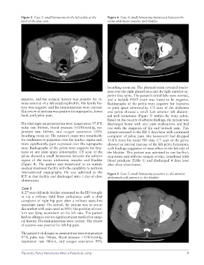
Figure 3  Case 3: small hematoma in the left pelvis at the   Figure 4  Case 4: small hematoma interposed between the
              level of the iliac vein.                           rectus abdominis muscles and bladder.


                                                                 breathing room air. The physical exam revealed tender-
                                                                 ness over the right gluteal area and the right anterior su-
                                                                 perior iliac spine. The patient’s initial labs were normal,
              negative, and his surgical history was positive for re-  and a bedside FAST exam was found to be negative.
              mote removal of a left-sided nephrolith. His family his-  Radiographs of the pelvis were negative for fractures
              tory was negative and his immunizations were current.   or joint space abnormality. CT scan of the abdomen
              His review of systems was positive for suprapubic, lower   and pelvis showed a small 2cm anterior left abdomi-
              back, and pelvic pain.                             nal wall hematoma (Figure 5) within the bony pelvis.
                                                                 Based on the paucity of adverse findings, the patient was
              His vital signs on presentation were temperature 97.8ºF,   discharged home with oral pain medications and bed
              pulse rate 86/min, blood pressure 141/81mmHg, res-  rest  with  the  diagnosis  of  hip  and  buttock  pain.  This
              piration rate 16/min, and oxygen saturation 100%   patient returned to the ED 3 days later with continued
              breathing room air. The patient’s exam was remarkable   complaint of pelvic pain. His hematocrit had dropped
              for tenderness to palpation over the lumbar region and,   11.8% from his initial ED visit. CT scan of the pelvis
              more significantly, pain expressed over the suprapubic   showed an interval increase of the left pelvic hematoma
              area. Radiographs of the pelvis were negative for frac-  with findings suggestive of mass effect on the left side of
              tures  or any  joint space  abnormality. CT  scan  of the   the bladder. This patient was admitted to our facility’s
              pelvis showed a small hematoma between the inferior   step-down unit with the surgery service, transfused with
              aspect of  the rectus abdominis  muscles and bladder   blood products (Table 1) and discharged 4 days later
              (Figure 4). The patient was transferred to an outside   after close observation.
              medical treatment facility with the capability to perform
              interventional  angiography.  He  was  admitted  to  the   Figure 5  Case 5: small hematoma posterior to the anterior
              ICU at that facility and discharged after 1 day of close   abdominal wall anterior to the bladder.
              observation.

              Case 5
              A 27-year-old male Soldier presented to the ED brought
              in via a military field litter ambulance with a chief
              complaint of right hip pain after a military static-line
              parachute jump. On arrival, the patient was in severe
              discomfort with pain rated as 8/10. His position of com-
              fort was lying recumbent on his left side. The patient
              had no allergies and no significant past medical or surgi-
              cal history. His immunizations were current. His review
              of systems was positive for left hip pain.

              The patient’s vital signs on presentation were temperature
              97ºF, pulse rate 78/min, blood pressure 137/83mmHg,
              respiration rate 18/min, and oxygen saturation  99%


              Traumatic Pelvic Hematoma After a Parachute Jump                                                 3