
Page 68 - Journal of Special Operations Medicine - Summer 2014
P. 68
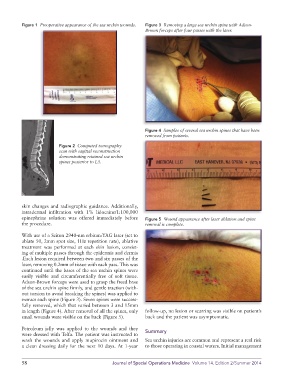
Figure 1  Preoperative appearance of the sea urchin wounds.  Figure 3  Removing a large sea urchin spine with Adson-
                                                             Brown forceps after four passes with the laser.


                                                             Figure 4  Samples of several sea urchin spines that have been
                                                             removed from patients.

                         Figure 2  Computed tomography
                         scan with sagittal reconstruction
                         demonstrating retained sea urchin
                         spines posterior to L5.


          skin changes and radiographic guidance. Additionally,
          intradermal infiltration with 1% lidocaine/1:100,000
          epinephrine  solution  was  offered  immediately  before   Figure 5  Wound appearance after laser ablation and spine
          the procedure.                                     removal is complete.

          With use of a Sciton 2940-nm erbium:YAG laser (set to
          ablate 50, 2mm spot size, 1Hz repetition rate), ablative
          treatment was performed at each skin lesion, consist-
          ing of multiple passes through the epidermis and dermis
          .Each lesion required between two and six passes of the
          laser, removing 0.2mm of tissue with each pass. This was
          continued until the bases of the sea urchin spines were
          easily  visible  and  circumferentially  free  of  soft  tissue.
          Adson-Brown forceps were used to grasp the freed base
          of the sea urchin spine firmly, and gentle traction (with-
          out torsion to avoid breaking the spines) was applied to
          extract each spine (Figure 3). Seven spines were success-
          fully removed, which that varied between 3 and 15mm
          in length (Figure 4). After removal of all the spines, only   follow-up, no lesion or scarring was visible on patient’s
          small wounds were visible on the back (Figure 5).  back and the patient was asymptomatic.

          Petroleum jelly was applied to the wounds and they
          were dressed with Telfa. The patient was instructed to   Summary
          wash the wounds and apply mupirocin ointment and   Sea urchin injuries are common and represent a real risk
          a clean dressing daily for the next 10 days. At 1-year   to those operating in coastal waters. Initial  management


          58                                   Journal of Special Operations Medicine  Volume 14, Edition 2/Summer 2014