
Page 207 - 2022 Ranger Medic Handbook
P. 207
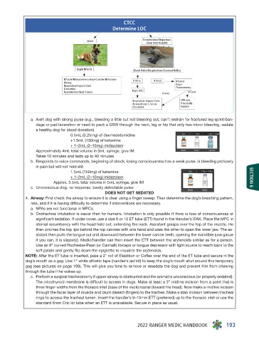
CTCC
                                   Determine LOC


          a.  Alert dog with strong pulse (e.g., bleeding a little but not bleeding out, can’t restrain for fractured leg splint/ban-
            dage or pad laceration or need to pack a GSW through the neck, leg or hip that only has minor bleeding, sedate
            a healthy dog for blood donation)
                       0.5mL (0.25mg) of dexmedetomidine
                       +1.5mL (150mg) of ketamine
                       + 1–2mL (2–10mg) midazolam
            Approximately 4mL total volume in 5mL syringe, give IM.
            Takes 10 minutes and lasts up to 40 minutes
          b.  Responds to voice commands, beginning of shock, losing consciousness has a weak pulse, is bleeding profusely
            in pain but will not hold still
                       1.5mL (150mg) of ketamine
                       + 1–2mL (2–10mg) midazolam                            SECTION 6
                 Approx. 3.5mL total volume in 5mL syringe, give IM
          c.  Unconscious dog, no response, barely detectable pulse
                                 DOES NOT GET SEDATED
         4.  Airway: First check the airway to ensure it is clear using a finger sweep. Then determine the dog’s breathing pattern,
          rate, and if it is having difficulty to determine if interventions are necessary.
          a.  NPAs are not functional in MPCs.
          b.  Orotracheal intubation is easier than for humans. Intubation is only possible if there is loss of consciousness or
            significant sedation. If under cover, use a size 9 or 10 ET tube (ETT) found in the Handler’s IFAK. Place the MPC in
            sternal recumbency with the head held out, extending the neck. Assistant grasps over the top of the muzzle. He
            then pinches the top lips behind the top canines with one hand and uses the other to open the lower jaw. The as-
            sistant then pulls the tongue out and downward between the lower canine teeth, opening the mandible (use gauze
            if you can, it is slippery). Medic/handler can then insert the ETT between the arytenoids similar as for a person.
            Use an 8" curved Rochester-Pean (or Carmalt) forceps or tongue depressor with light source to reach back to the
            soft palate and gently flip down the epiglottis to visualize the arytenoids.
         NOTE: After the ET tube is inserted, pass a 2" roll of Elastikon or Coflex over the end of the ET tube and secure in the
         dog’s mouth as a gag. Use 1" white athletic tape (handler’s aid kit) to keep the dog’s mouth shut around this temporary
         gag (see pictures on page 198). This will give you time to remove or resedate the dog and prevent him from chewing
         through the tube if he wakes up.
          c.  Perform a surgical tracheostomy if upper airway is obstructed and the animal is unconscious (or properly sedated).
            The cricothyroid membrane is difficult to access in dogs. Make at least a 3” midline incision from a point that is
            three finger widths from the thoracic inlet (base of the neck) rostral (toward the head). Now make a midline incision
            through the facial layer of muscle and blunt dissect (fingers) to the trachea. Make a stab incision between tracheal
            rings to access the tracheal lumen. Insert the handler’s 9–10mm ETT (preferred) up to the thoracic inlet or use the
            standard 6mm Cric kit tube when an ETT is unavailable. Secure in place as usual.


                                           2022 RANGER MEDIC HANDBOOK  193