
Page 62 - JSOM Winter 2024
P. 62
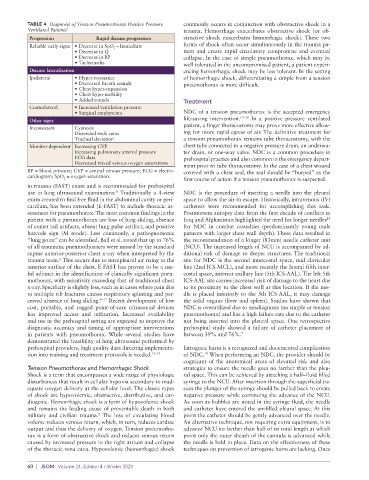
TABLE 4  Diagnosis of Tension Pneumothorax Positive Pressure   commonly occurs in conjunction with obstructive shock in a
          Ventilated Patients 5                              trauma.  Hemorrhage  exacerbates  obstructive  shock  (or  ob-
           Progression           Rapid disease progression   structive  shock  exacerbates  hemorrhagic  shock).  These  two
           Reliable early signs • Decrease in SpO – Immediate  forms of shock often occur simultaneously in the trauma pa-
                                      2
                         • Decrease in Q                     tient  and create  rapid  circulatory  compromise  and eventual
                         • Decrease in BP                    collapse. In the case of simple pneumothorax, which may be
                         • Tachycardia                       well tolerated in the uncompromised patient, a patient experi-
           Disease lateralization                            encing hemorrhagic shock may be less tolerant. In the setting
           Ipsilateral   • Hyper-resonance                   of hemorrhagic shock, differentiating a simple from a tension
                         • Decreased breath sounds           pneumothorax is more difficult.
                         • Chest hyper-expansion
                         • Chest hypo-mobility
                         • Added sounds                      Treatment
           Contralateral   • Increased ventilation pressure
                         • Surgical emphysema                NDC of a tension pneumothorax is the accepted emergency
           Other signs                                       life-saving intervention. 15–18  In a positive pressure ventilated
           Inconsistent  Cyanosis                            patient, a finger thoracostomy may prove more effective allow-
                         Distended neck veins                ing for more rapid egress of air. The definitive treatment for
                         Tracheal deviation                  a tension pneumothorax remains tube thoracostomy, with the
           Monitor-dependent Increasing CVP                  chest tube connected to a negative pressure drain, an underwa-
                         Increasing pulmonary arterial pressure  ter drain, or one-way valve. NDC is a common procedure in
                         ECG data                            prehospital practice and also common in the emergency depart-
                         Decreased mixed venous oxygen saturations
                                                             ment prior to tube thoracostomy. In the case of a chest wound
          BP = blood pressure; CVP = central venous pressure; ECG = electro-  covered with a chest seal, the seal should be “burped” as the
          cardiogram; SpO  = oxygen saturation.
                      2                                      first course of action if a tension pneumothorax is suspected.
          in trauma (FAST) exam and is recommended for prehospital
          use in lung ultrasound examination.  Traditionally a 4-view   NDC is the procedure of inserting a needle into the pleural
                                       8
          exam created to find free fluid in the abdominal cavity or peri-  space to allow the air to escape. Historically, intravenous (IV)
          cardium, has been extended (E-FAST) to include thoracic as-  catheters were recommended for accomplishing this task.
          sessment for pneumothorax. The most common findings in the   Postmortem autopsy data from the first decade of conflicts in
                                                                                                            14
          patient with a pneumothorax are loss of lung sliding, absence   Iraq and Afghanistan highlighted the need for longer needles
          of comet tail artifacts, absent lung pulse artifact, and positive   for NDC in combat casualties (predominantly young male
          barcode sign (M mode). Less commonly, a pathognomonic   patients with larger chest wall depth). These data resulted in
          “lung point” can be identified. Ball et al. noted that up to 76%   the recommendation of a longer (83mm) needle catheter unit
          of all traumatic pneumothoraces were missed by the standard   (NCU). The increased length of NCU is accompanied by ad-
          supine anterior-posterior chest x-ray when interpreted by the   ditional risk of damage to deeper structures. The traditional
                    6
          trauma team.  This occurs due to intrapleural air rising to the   site for NDC is the second intercostal space, mid clavicular
          anterior surface of the chest. E-FAST has proven to be a use-  line (2nd ICS-MCL), and more recently the lateral fifth inter-
          ful adjunct in the identification of clinically significant pneu-  costal space, anterior axillary line (5th ICS-AAL). The left 5th
          mothorax, with sensitivity exceeding that of traditional chest   ICS-AAL site carries increased risk of damage to the heart due
          x-ray. Specificity is slightly less, such as in cases where pain due   to its proximity to the chest wall at this location. If the nee-
          to multiple rib fractures causes respiratory splinting and per-  dle is placed inferiorly to the 5th ICS-AAL, it may damage
          ceived absence of lung sliding. 8–11  Recent development of low   the solid organs (liver and spleen). Studies have shown that
          cost, portable, and rugged point-of-care ultrasound devices   NDC is overutilized due to misdiagnosis (no simple or tension
          has improved access and utilization. Increased availability   pneumothorax) and has a high failure rate due to the catheter
          and use in the prehospital setting are expected to improve the   not being inserted into the pleural space. One retrospective
          diagnostic accuracy and timing of appropriate interventions   prehospital study showed a failure of catheter placement of
          in patients with pneumothorax.  While several studies have   between 39% and 76%. 7
          demonstrated the feasibility of lung ultrasound performed by
          prehospital providers, high quality data directing implementa-  Iatrogenic harm is a recognized and documented complication
          tion into training and treatment protocols is needed. 12–14  of NDC.  When performing an NDC, the provider should be
                                                                    19
                                                             cognizant of the anatomical areas of elevated risk and also
          Tension Pneumothorax and Hemorrhagic Shock         strategies to ensure the needle goes no farther than the pleu-
          Shock is a term that encompasses a wide range of physiologic   ral space. This can be achieved by attaching a half–fluid filled
          disturbances that result in cellular hypoxia secondary to inad-  syringe to the NCU. After insertion through the superficial tis-
          equate oxygen delivery at the cellular level. The classic types   sues the plunger of the syringe should be pulled back to create
          of shock are hypovolemic, obstructive, distributive, and car-  negative pressure while continuing the advance of the NCU.
          diogenic. Hemorrhagic shock is a form of hypovolemic shock   As soon as bubbles are noted in the syringe fluid, the needle
          and remains the leading cause of preventable death in both   and catheter have entered the air-filled pleural space. At this
                                 2
          military and civilian trauma.  The loss of circulating blood   point the catheter should be gently advanced over the needle.
          volume reduces venous return, which, in turn, reduces cardiac   An alternative technique, not requiring extra equipment, is to
          output and thus the delivery of oxygen. Tension pneumotho-  advance NCU no farther than half of its total length at which
          rax is a form of obstructive shock and reduces venous return   point only the outer sheath of the cannula is advanced while
          caused by increased pressure in the right atrium and collapse   the needle is held in place. Data on the effectiveness of these
          of the thoracic vena cava. Hypovolemic (hemorrhagic) shock   techniques on prevention of iatrogenic harm are lacking. Once

          60  |  JSOM   Volume 24, Edition 4 / Winter 2024