
Page 12 - JSOM Summer 2023
P. 12
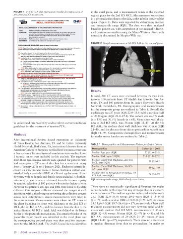
FIGURE 1  TCCC CLS skill instruction: Needle decompression of   in the axial plane, and a measurement taken in the matched
          the chest (NDC) instruction.                       sagittal plane for the 2nd ICS MCL. Measurements were taken
                                                             in a perpendicular plane to the skin, at the inferior border of the
                                                             space (Figure 2). Data were reported by determining median
                                                             and interquartile range (IQR). The data were then analyzed
                                                             based on patient sex, with comparison of non- normally distrib-
                                                             uted continuous variables using the Mann-Whitney U test, with
                                                             normality determined by Shapiro-Wilk test.

                                                             FIGURE 2  Sample measurement at 5th ICS AAL in the coronal plane.


                                                             Results

                                                             In total, 200 CT scans were reviewed between the two insti-
                                                             tutions: 100 patients from UT Health San Antonio, San An-
                                                             tonio, TX and 100 patients from St. Luke’s University Health
                                                             Network, Bethlehem, PA. Demographics  and  measurements
                                                             for the composite group are outlined in Table 1. Overall, the
                                                             median age was 27 years (IQR 22.0–33.8) with a median BMI
                                                             of 23.8 kg/m  (IQR 21.8–27.2). The cohort was 69.5% male
                                                                       2
                                                             (n = 139) and 30.5% female (n = 61). Mean chest wall thick-
          to understand this possibility and to inform current and future   ness at 2nd ICS MCL was 38-mm (IQR 32–45). At the 5th
          guidelines for the treatment of tension PTX.       ICS AAL, the median chest wall thickness was 30-mm (IQR
                                                             21–40), and the distance from skin to pericardium was 66-mm
                                                             (IQR 54–79). Comparative demographics and measurements
          Methods
                                                             for males versus females are outlined in Table 2.
          After Institutional Review Board exemption at University
          of Texas Health, San Antonio, TX and St. Lukes University   TABLE 1  Demographic and Measurement Values for Entire Cohort
          Health Network, Bethlehem, PA, institutional datasets from an
          American College of Surgeons verified level I trauma center and   Demographics       Cohort (n = 200)
          a Pennsylvania Trauma System Foundation state-verified Level   Median Age, year (IQR)  27 (22.0–33.8)
          1 trauma center were included in this analysis. The registries   Median BMI, kg/m  (IQR)  23.8 (21.8–27.2)
                                                                           2
          from these two trauma centers were queried for patients who   Median Chest Wall Thickness, 2nd ICS   38 (32–45)
          had undergone a CT scan of the chest for a traumatic injury   MCL, mm (IQR)
          from 1 January 2016 to 1 January 2021. Exclusion criteria in-  Median Chest Wall Thickness, 5th ICS   30 (21–40)
          cluded an intrathoracic mass or lesion. Inclusion criteria con-  AAL, mm (IQR)
          sisted of body mass index (BMI) of ≤30 and age between 18 and   Median Skin to Pericardium Distance, 5th   66 (54–79)
          40 years, with both male and female sexes included. At both in-  ICS AAL, mm (IQR)
          stitutions, patient data were obtained from the trauma registry   IQR = interquartile range, BMI = body mass index.
          by random selection of 100 patients who met inclusion criteria.
          However the patient’s sex, age, and BMI were blind to the data   There were no statistically significant differences for males
          collector. One surgeon collector reviewed the images at each   versus females with respect to any demographic or measure-
          institution with a third surgeon reviewing the collection process   ment parameter. The median ages for males and females were
          across both institutions to ensure measurements were taken in   26.0 (IQR 22.0–34.0) versus 29.0 years (IQR 21.5–34.0)
          the same manner. Measurements were taken on CT scans of   (p = .76) with a median BMI of 24.0 (IQR 21.5–27.4) versus
          the chest including the chest wall thickness at the 2nd ICS at   23.5 kg/m  (IQR 19.7–26.6) (p =.17), respectively. Chest wall
                                                                     2
          MCL, the 5th ICS at AAL, and the distance from the skin to the   thickness measurements did not vary between males and fe-
          pericardium at 5th ICS at AAL. AAL was defined as the lateral   males with median 2nd ICS MCL measurements of 39-mm
          border of the pectoralis musculature. The anterior border of the   (IQR 32–45) versus 38-mm (IQR 32–47) (p =.45) and 5th
          pectoralis major muscle was identified in the axial plane and   ICS AAL measurements of 29 (IQR 21–38) versus 34-mm
          the corresponding coronal plane was then used for measure-  (IQR 23–43) (p =.07), respectively. There were no differences
          ment for the 5th ICS AAL. The 2nd ICS was likewise identified   in median distances from skin to pericardium for males or

          10  |  JSOM   Volume 23, Edition 2 / Summer 2023