
Page 118 - JSOM Summer 2023
P. 118
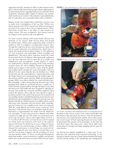
supportive and SpO  remained at 100% for the duration of sur-  FIGURE 3  Tissue debridement of right leg and vessel ligation.
                         2
          gery. Cardiovascular status had required volume replacement as
          an intervention, but he responded well and was stabilized. Body
          temperature remained stable. Depth of sedation was continu-
          ously monitored by trended vitals, stimulus to procedural pain,
          lack of a gag reflex, and occasional corneal reflex evaluation.

          Surgical towels were draped above and below operative sites
          to enable sterile manipulation of the leg. After 18D(a) com-
          pleted surgical scrub, 18D(c) placed loose bilateral tourniquets
          proximal to the surgical sites. Using a surgical marker, 18D(a)
          marked the incision lines in the shape of a fish mouth to fa-
          cilitate closure. The team conducted a final timeout and the
          tourniquets on the operative side was tightened.

          An initial incision utilizing a #10 scalpel blade followed skin
          markings on the anterior thigh moving down and around
          the lateral aspect. 18D(a) and a supporting medic worked in
          tandem in order to complete a circumferential incision. Once
          through the epidermis, dermis, and subcutaneous tissue, blunt
          and iris scissor dissection was performed to retract tissue to
          expose the muscle and fascial planes (Figure 3). The saphenous
          vein was superficial and incidentally nicked providing easy
          identification. This led to blunt dissection around the vessel
          for proximal site for silk ligation. After ligating the saphenous
          vein, the long tails were not cut from the tie to enable easy   FIGURE 4  Bone saw use to complete the amputation of the right leg.
          identification  and  manipulation.  Further  ligation  of  vessels
          was performed by complete wrapping of the vessel and three
          surgeon’s knots for secure holding. Transection through the
          muscle groups began on the anterior aspect with the quadri-
          ceps tendon, down through the vastus intermedius. This was
          followed by the vastus lateralis into the iliotibial tract. With
          the leg elevated, the semitendinosus, semimembranosus, and
          the biceps femoris were transected into the medial aspect for
          the sartorius and the vastus medialis. While navigating muscle
          groups, 18D(a) was able to identify the vessel and nerve bun-
          dles, including a visibly strong pulsating femoral artery. After
          exposing these vessels at a proximal location, a needle on 5-0
          Vicryl inserted perpendicularly through the femoral arty, then
          the suture tails and needle end were wrapped in opposing di-
          rections. The needle was removed, and three surgeon’s knots
          secured  the  ligation.  Next,  silk  ties  were  placed  to  ligate  a
          proximal portion of the vessel. This process was repeated with
          surrounding vessels as needed leaving long suture tails for easy
          identification and manipulation. Long nerve endings were also
          ligated with silk ties. Distal traction was pulled, and the vessels
          and nerves were transected with iris scissors. After vessel liga-
          tion and transection was complete excess tissue was removed.
          With the vessels ligated and muscle groups transected, blunt
          dissection of the muscle tissue was performed from the prox-
          imal femur to approximately five inches from the distal fem-  site. A pen rose drain was sutured in place, securing the top of
          oral epicondyle. A bone rasp was used to identify and isolate   the drain to the interior aspect of the vastus intermedius and
          a location for gigli saw use. The femur was transected, and a   the bottom of the opening to the interior of the biceps femoris.
          sterile rasp was used to remove sharp edges (Figure 4). Next, a   Dermal tissue was approximated, and the fish mouth incision
          ¼-in sterilized drill bit and a commercial power drill wrapped   was closed using 3-0 Vicryl ensuring the pen rose drain would
          in a sterile towel was utilized to drill a hole on the posterior   be functional. The entirety of the procedure on the right leg
          aspect of the femur for anchoring. As a test of ligation efficacy   took three hours. The team took a short break with visual
          and for insight into potential signs of tissue viability, 18D(c)   monitoring of vital signs and began prepping the left leg in the
          released the tourniquets; no hemorrhage was noted and tis-  same manner.
          sue rapidly regained color. Four liters of normal saline were
          used for low pressure irrigation and all remaining soft tissue   The left leg was greatly simplified by a lesser need for ex-
          was also deemed viable. 2-0 Vicryl was threaded through the   ploration and expedited with improved knowledge of the
          quadriceps tendon, vastus intermedius, medialis, and lateralis,     procedure-specific anatomy. A large abscess between the femur
          which were anchored to the previously drilled femoral anchor   and the muscle groups of the quadriceps was unexpectedly

          116  |  JSOM   Volume 23, Edition 2 / Summer 2023