
Page 44 - JSOM Winter 2022
P. 44
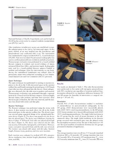
FIGURE 1  Reactor device.


                                                             FIGURE 2  Reactor
                                                             technique.


          National Institute of Health. Experiments were performed on
          30–40 kg Sus scrofa swine by surgical residents (postgraduate
          year [PGY]-2 and 3).
          After intubation, intrathoracic access was established via nee-
          dle catheterization in the 2nd or 3rd intercostal space. At this
          point, 600mL of air was instilled into the hemithorax and
          pneumothorax was confirmed with x-ray.  The intervention
          was randomized to surgical resident (PGY-2, -3) and technique
          (OT, RT). Time was recorded. Pos-procedural radiography was
          used to confirm pneumothorax resolution and tube placement.
          Thoracoscopic evaluation was performed by a board-certified                      FIGURE 3 Reactor
          thoracic surgeon to evaluate and record evidence of injury,                      technique.
          estimated blood loss (EBL), and incision length. Radiographs
          were read by a board-certified thoracic surgeon blinded to
          resident and technique. After five interventions on one hemi-
          thorax, the contralateral hemithorax was utilized.  After 10
          procedures, swine were euthanized according to local Institu-
          tional Animal Care and Use Committee (IACUC) protocol.
          Open Technique
          The open technique was performed by making an incision su-  Results
          perior to the rib in the 4–6th intercostal space in the anterior
          axillary line and bluntly entering the pleural space. A 28-French   The results are depicted in Table 1. Fifty tube thoracostomies
          chest tube was then advanced into the thorax. Chest radiogra-  were performed on five swine with iatrogenic pneumothorax
          phy was obtained after the tube was in position and secured.   with 98% resolution rate regardless of insertion method. Ran-
          Thoracoscopic evaluation was performed through a separate   domization allowed for no significant difference between OT
          incision by a board-certified thoracic surgeon. Incision length,   and RT groups for laterality of intervention or surgical resi-
          thoracic injury, tube position, and time were recorded. After   dent (p = .89 and .41).
          thoracoscopic evaluation, the tube was removed, and the inci-
          sion was closed with suture and skin glue.         Resolution
                                                             Overall, 96% of tube thoracostomies resulted in resolution
          Reactor Technique                                  of pneumothorax on post-procedural radiography. In the
          The Reactor technique was performed using the device with   OT group, 24/25 thoracostomies were successfully placed,
          sleeve. A skin incision was made above the rib in the 4–6th   with 24/25 (96%) resolving pneumothorax. In the RT group,
          intercostal space in the anterior axillary line. The pleural space   25/25 thoracostomies were successfully performed, with 24/25
          was entered using sequential firings of the spring-loaded Re-  (96%) resolving pneumothorax. The single failed resolution in
          actor device (Figure 2). The sleeve was passed over the device   the OT group was the result of errant placement in the sub-
          into the pleural space. The device was withdrawn, leaving the   cutaneous tissue. The single failed resolution in the Reactor
          sleeve in place. A 28-French chest tube was advanced through   group occurred with successful entry into the pleural cavity
          the sleeve (Figure 3). The sleeve was removed and the tube   but incomplete evacuation of air. The size of this study (n = 50)
          was secured, followed by radiographic confirmation. Thoraco-  and the equal numbers of resolution between the two groups
          scopic evaluation was performed, and the tube was removed.   prevented evaluation of significance.
          The incision was closed with suture and skin glue. Incision
          length, thoracic injury, position, and time were recorded.  Time
                                                             The average insertion time overall was 37.9 seconds (standard
          Each insertion was evaluated by method (OT, RT), pneumo-  deviation [SD] ± 14 seconds). OT average insertion time was
          thorax resolution, time, incision length, placement, complica-  38.2 seconds (SD ± 10 seconds). OT maximum time (T max ) was
          tions, and EBL.                                    63.8 seconds; minimum time (T ) was 19.3 seconds. This was
                                                                                     min
          42  |  JSOM   Volume 22, Edition 4 / Winter 2022