
Page 47 - 2022 Spring JSOM
P. 47
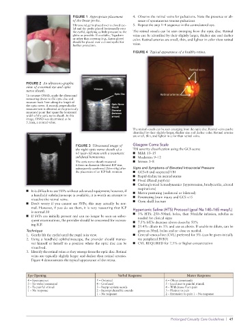
FIGURE 1  Appropriate placement   4.  Observe the retinal veins for pulsations. Note the presence or ab-
                                    of the linear probe.           sence of spontaneous venous pulsations
                                    Ultrasound gel is placed over a closed eye-  5.  Repeat the step 1–4 sequence in the contralateral eye.
                                    lid and the probe placed horizontally over
                                    the eyelid, applying as little pressure to the   The retinal vessels can be seen emerging from the optic disc. Retinal
                                    globe as possible. If available, Tegaderm   veins can be identified by their slightly larger, thicker size and darker
                                    or other thin covering (e.g., Latex glove)   color. Retinal arteries are small, thin, and lighter in color than retinal
                                    should be placed over a closed eyelid for
                                    further protection.          veins.
                                                                 FIGURE 4  Typical appearance of a healthy retina.


              FIGURE 2  An ultrasono-graphic
              view of a normal eye and optic
              nerve sheath.
              To measure ONSD, apply the ultrasound
              measuring device to the optic disc and
              measure back 3mm along the length of
              the optic nerve. A second, perpendicular
              measurement is obtained at the previously
              measured point that spans the horizontal
              width of the optic nerve sheath. In this
              image, ONSD was determined to be
              5.1mm, a normal value.
                                                                 The retinal vessels can be seen emerging from the optic disc. Retinal veins can be
                                                                 identified by their slightly larger, thicker size and darker color. Retinal arteries
                                                                 are small, thin, and lighter in color than retinal veins.

                                     FIGURE 3  Ultrasound image of   Glasgow Coma Scale
                                     the right optic nerve sheath of a   TBI severity classification using the GCS score:
                                     61-year-old man with a traumatic   ■   Mild: 13–15
                                     subdural hematoma.          ■   Moderate: 9–12
                                     The optic nerve sheath measured   ■   Severe: 3–8
                                     6.8mm in diameter. Elevated ICP was
                                     subsequently confirmed (26mmHg) after   Signs and Symptoms of Elevated Intracranial Pressure
                                     the placement of an ICP bolt monitor.  ■   GCS<8 and suspected TBI
                                                                 ■   Rapid decline in mental status
                                                                 ■   Fixed dilated pupils(s)
                                                                 ■   Cushing’s triad hemodynamics (hypertension, bradycardia, altered
                                                                   respirations)
              ■   It is difficult to see SVPs without advanced equipment; however, if   ■   Motor posturing (unilateral or bilateral)
                a handheld ophthalmoscope is available, it is worth an attempt to   ■   Penetrating brain injury and GCS <15
                visualize the retinal veins.
              ■   Don’t  worry  if  you  cannot  see  SVPs;  this  may  actually  be  nor-  ■   Open skull fracture
                mal. However, if you do see them, it is very reassuring that ICP   Hypertonic Saline (HTS) Protocol (goal Na 140–165 meq/L)
                is normal.10
              ■   If SVPs are initially present and can no longer be seen on subse-  ■   3% HTS: 250–500mL bolus, then 50mL/hr infusion, rebolus as
                                                                   needed for clinical signs
                quent examinations, the provider should be concerned for increas-  ■   7.5% HTS: decrease above doses by 50%
                ing ICP.
                                                                 ■   23.4%: dilute to 3% and use as above. If unable to dilute, can be
              Technique                                            given as 30mL bolus and re- dose as needed.
              1.  Gently lift the eyelid until the pupil is in view.  ■   Central venous line (CVL) preferred for 3% (can be given initially
              2.  Using a handheld ophthalmoscope, the provider should maneu-  via peripheral IV/IO)
                ver himself or herself to a position where the optic disc can be   ■   CVL REQUIRED for 7.5% or higher concentration
                visualized.
              3.  Identify the retinal veins as they emerge from the optic disc. Retinal
                veins are typically slightly larger and darker than retinal arteries.
                Figure 4 demonstrates the typical appearance of the retina.


              Eye Opening                                Verbal Response                  Motor Response
              4 – Spontaneous                  5 – Oriented                     6 – Obeys commands
              3 – To verbal command            4 – Confused                     5 – Localizes to painful stimuli
              2 – To painful stimuli           3 – Inappropriate words          4 – Withdraws from pain
              1 – No response                  2 – Incomprehensible sounds      3 – Flexion to pain
                                               1 – No response                  2 – Extension to pain 1 – No response


                                                                                   Prolonged Casualty Care Guidelines  |  45