
Page 115 - 2020 JSOM Winter
P. 115
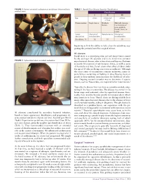
FIGURE 1 Patient on initial evaluation at an African Union military   TABLE 1  Buruli Ulcer Disease Severity Classification
              medical clinic.                                                             Classification
                                                                                  I         II           III
                                                                 Size           <5cm      >5cm and     >15cm
                                                                                           <15cm
                                                                 Dissemination  Single lesion  Multiple   Multiple lesions;
                                                                                        lesions    involvement of
                                                                                                   eyes, genitals,
                                                                                                   breast, bone, or
                                                                                                   joints
                                                                 Lesion      Ulcerative/  Nonulcerative Ulcerative/
                                                                 description  nonulcerative        nonulcerative
                                                                 Prevalence, %   32         35           33


                                                                 beginning to limit his ability to fully close the palpebrae, sug-
                                                                 gesting the potential need for surgical release.

                                                                 M. ulcerans
                                                                 Buruli ulcer is a necrotizing skin and soft tissue lesion caused
                                                                 by the pathogen M. ulcerans and is the third most common
              FIGURE 2  Infraorbital ulcer on initial evaluation.  mycobacterial disease, after tuberculosis and leprosy. Endemic
                                                                 to nearly 30 countries of sub-Saharan Africa, as well as parts
                                                                 of Australia and Asia, Buruli ulcers often affect children under
                                                                 the age of 15 who are living near rivers or wetlands.  Although
                                                                                                        1
                                                                 the exact cause of transmission remains unknown, many ex-
                                                                 perts believe swimming or bathing in slow-flowing rivers or
                                                                 ponds in these endemic areas increases the likelihood of infec-
                                                                 tion. Ongoing research is under way to determine if aquatic
                                                                 insects, such as Naucoridae, are responsible for transmission. 2

                                                                 Typically, the disease first manifests as a painless nodule, origi-
                                                                 nating in the face or extremities. The disease may remain in its
                                                                 latent stage until activated, often by superficial trauma. Even-
                                                                 tually, these nodules become poorly demarcated ulcers which
                                                                                  3
                                                                 may penetrate to bone.  Buruli ulcers are distinguishable from
                                                                 many other mycobacterial diseases due to the absence of fever
                                                                 and lymphadenopathy, aiding in diagnosis. Though classically
                                                                 described as a painless lesion, our experience with this pa-
                                                                 tient’s lesions being painful is consistent with numerous recent
                                                                       4–7
                                                                 reports.  Bacterial superinfection may cause fever, as in this
              M. ulcerans  complicated by secondary bacterial infection,   case, and cloud the clinical diagnosis. Polymerase chain reac-
              based on lesion appearance, distribution, and progression de-  tion testing using a punch biopsy shows the highest sensitivity
              spite atypical symptoms of pain and fever. According to World   and specificity of available laboratory testing, both of which
              Health Organization guidelines, this patient had Class III Bu-  approach 100%. In the resource-limited environment, diag-
                                                                              8
              ruli ulcer disease, given the number and distribution of ulcers   nosis is made based on patient history, presentation, and phys-
              (Table 1). They recommended treatment with an extended   ical exam. The prognosis is generally favorable if diagnosed
              course of clarithromycin and rifampin, but neither was avail-  and treated promptly but may lead to lifelong debilitation if
              able in the austere environment. We substituted azithromycin   left untreated. 9,10  Studies in Ghana and Benin have shown ex-
              and requisitioned rifampin. When the patient returned after 2   tensive physical, psychological, and social impairments as a
              weeks of azithromycin, his ulcers had progressed. We sought   result of this disease. 11–14
              further consultation, and both specialists recommended add-
              ing ciprofloxacin to the regimen.
                                                                 Surgical Treatment
              At the next follow-up, the ulcers had not progressed further.   Patient selection for surgery parallels the management of other
              At  that  time,  we had  received  a supply  of  rifampin  and  he   infected wounds. If local bedside debridement cannot cleanse
              was started on a regimen of rifampin, ciprofloxacin, and azi-  the wounds of purulence and adequately assess whether in-
              thromycin. We initiated a 12-week antibiotic course due to   fection extends into subcutaneous pockets, the patient should
              the fastidiousness of M. ulcerans, but unfortunately the pa-  be considered for operative debridement. As this is primarily
              tient was temporarily lost to follow-up after 10 weeks. Five   a medically managed disease due to the discovery of success-
              months later, he presented again with worsening lesions. He   ful antibiotic regimens, skin grafting should be reserved for
              subsequently completed a new 12-week course of triple antibi-  patients with nonhealing, but noninfected, lesions that have
              otics with resulting slow progressive healing of the old lesions,   significant functional impact. These include lesions over major
              which we interpreted as a clinical sign of successful treatment.   joints and in the periorbital region that can affect eye closure,
              Scarring of the ulceration below his left eye, however, was   as occurred in our patient. Skin grafting requires specialized

                                                                                Unusual Tropical Disease in East Africa  |  113