
Page 76 - JSOM Winter 2017
P. 76
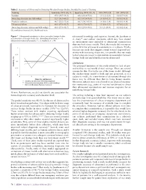
Table 2  Accuracy of Ultrasound in Detecting Wooden Foreign Bodies, Stratified by Level of Training
           Training                         Sensitivity (95% CI), %  Specificity (95% CI), %  LR+ (95% CI)  LR− (95% CI)
           Resident                           47.0 (39.7–54.5)   68.6 (61.4–75.1)  1.5 (1.2–2.0)  0.8 (0.7–0.9)
           Attending physician (no fellowship)  52.5 (36.3–68.2)  62.5 (45.8–76.8)  1.4 (0.9–2.3)  0.8 (0.5–1.1)
           Physician assistant                46.7 (22.2–72.6)   66.7 (38.7–87.0)  1.4 (0.6–3.4)  0.8 (0.5–1.4)
           Ultrasound fellow                  60.0 (17.0–92.7)   60.0 (17.0–92.7)  1.5 (0.4–5.5)  0.7 (0.2–2.5)
           Attending physician (ultrasound fellowship)  60.0 (17.0–92.7)  80.0 (29.9–99.0)  3.0 (.5–19.9)  0.5 (0.2–1.6)
          CI, confidence interval; LR, likelihood ratio.

          Figure 1  Ultrasound sensitivity to detect wooden foreign bodies   ultrasound knowledge and expertise. Second, the Jacobson et
          as a function of foreign body size. Attending physicians, n = 8;   al. study  used cadaver specimens, which may have created
                                                                    20
          ultrasound-trained physicians, n = 2; midlevel, n = 40.
                                                             an environment more amenable to ultrasound examinations
                                                             than our fresh-tissue models. Third, their study did not impose
                                                             a time limit for ultrasound examinations on subjects. Finally,
                                                             because our study does suggest a trend toward improved sen-
                                                             sitivity with increasing object size, it is possible that our study
                                                             lacked adequate power to demonstrate an association between
                                                             foreign body size and identification via ultrasound.

                                                             Limitations
                                                             The principal limitation of this study related to lack of gen-
                                                             eralizability to real-world clinical settings. There are several
                                                             reasons for this. First is the use of the tissue model. Although
                                                             the chicken-tissue model is fresh and not preserved, as is a
                                                             cadaveric model, the transmission of ultrasound through this
                                                             tissue may be different than that in living human models.
                                                             Moreover, subjects were not allowed to use physical examina-
                                                             tion maneuvers (e.g., point of maximal tenderness) to direct
                                                             their ultrasound examinations and increase suspicion for an
                                                             underlying foreign body.
          10 mm. Furthermore, we did not identify any association be-
          tween diagnostic accuracy and education level.     The setting including a time limit imposed on our subjects
                                                             similarly may limit generalizability. Our intent was to simu-
          The pooled sensitivity was 48.4% for the use of ultrasound to   late the circumstances of a busy emergency department and
          detect wooden foreign bodies. This aligns with the lower range   consistently limit the resource of available time to complete
          of values previously reported in the literature for detection of   the procedure. However, had we offered subjects more time
          various foreign bodies in tissue models (50%  to 52.6% ).   to complete their examinations, it is possible they would have
                                                        6
                                               10
          These values contrast with studies reporting significantly   achieved higher diagnostic accuracy. On the other hand, to the
          higher sensitivities for ultrasound in detecting foreign bodies   point of simulating an emergency department environment,
          ranging up to 93% to 100%. 12,13,19  There are several potential   our subjects performed their examinations in a relatively
          explanations why other studies reported significantly higher   quiet, dark, and secluded room, which may have increased
          sensitivities. First, some of these studies entailed didactic ses-  their diagnostic accuracy and may not approximate the typi-
          sions immediately before data collection, which may have   cal clinical care setting in the emergency department.
          improved subject diagnostic accuracy.  Second, some used
                                         13
          differing tissue models, such as human cadaveric tissue, and it   Another limitation is the sample size. Though our dataset
          is possible that this medium is more amenable to sonographic   comprised 500 ultrasound studies, only 50 studies were per-
          penetration or creates more echogenic contrast between mod-  formed for each foreign body size. This yielded a study pow-
          eled tissue and foreign body than our fresh-tissue model. 12,19    ered to detect sensitivity differences of 20% or more. It may
          Third, none of these prior studies reported an imposed time   be that diagnostic accuracy, indeed, improves with increasing
          limit  on participants and  may  have enabled  more  time  for   foreign body size but in smaller increments, which could not
          aspects of procedure completion, increasing diagnostic test   be detected because of a small sample size.
          characteristics. Finally, some of these studies aggregated ex-
          aminations of tissue models with alternative substances (e.g.,   Future Research
          metal, glass), the inclusion of which may have confounded ac-  Our results highlight several potential areas for investigations.
          curacy estimates. 13,17                            An ideal investigation may be a clinical study of emergency de-
                                                             partment patients with suspected radiolucent foreign bodies in
          Our findings contrast with at least one study that suggested im-  penetrating wounds. Such a study would ideally compare the
          proved sensitivity of ultrasound for detecting wooden foreign   diagnostic accuracy of ultrasound as performed in the emer-
          bodies with increasing size.  That study, by Jacobson et al.,    gency department versus a gold standard such as magnetic res-
                               20
                                                         20
          reported a sensitivity of 86.7% for foreign bodies measuring   onance imaging or surgical exploration. Such a study would
          2.5mm and 93.3% for foreign bodies measuring 5.0mm. How-  likely be logistically challenging to accumulate sufficient num-
          ever, the subjects differed from our emergency medicine pro-  bers of patients for a well-powered analysis. In the interim,
          vider population as musculoskeletal radiologists with extensive   further insight may be gained from replication of our study

          74  |  JSOM   Volume 17, Edition 4/Winter 2017