
Page 96 - Journal of Special Operations Medicine - Winter 2015
P. 96
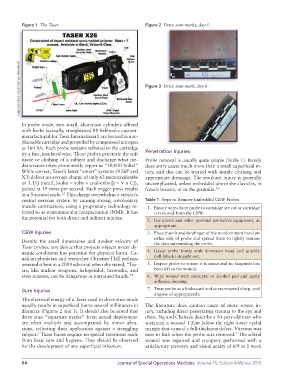
Figure 1  The Taser                                Figure 2  Drive stun marks, day 1.


         Image used with permission from Taser International  Figure 3  Drive stun mark, day 6.


          In probe mode, two small, aluminum cylinders affixed
          with barbs (actually, straightened #8 fishhooks custom-
          manufactured for Taser International) are housed in a re-
          placeable cartridge and propelled by compressed nitrogen
          at 160 ft/s. Each probe remains tethered to the cartridge
          by a fine, insulated wire. These probes penetrate the soft   Penetration Injuries
          tissue or clothing of a subject and discharge what me-  Probe removal is usually quite simple (Table 1). Rarely
          dia sources often prominently report as “50,000 Volts!”   does entry cause much more than a small superficial in-
          While correct, Taser’s latest “smart” systems (X26P and   jury, and this can be treated with simple cleaning and
          X3) deliver an average charge of only 63 microcoulombs   appropriate dressings. The resultant injury is generally
          or 3.15J (recall, joules = volts × coulombs [J = V × C]),   uncomplicated, unless embedded above the clavicles, in
          pulsed at 19 times per second. Each trigger press results   female breasts, or in the genitalia. 5,6
                          2,3
          in a 5-second cycle.  This charge overwhelms a subject’s
          central  nervous system by  causing  strong, involuntary   Table 1  Steps to Remove Embedded CEW Probes
          muscle contractions, using a proprietary technology re-  1.  Ensure wires from probe to cartridge are cut or cartridge
          ferred to as neuromuscular incapacitation (NMI). It has   is removed from the CEW.
          the potential for both direct and indirect injuries.
                                                              2.  Use gloves and other personal protective equipment, as
                                                                 appropriate.
          CEW Injuries                                        3.  Place thumb and forefinger of the nondominant hand on
                                                                 either side of probe and spread them to tightly tension
          Despite the small dimensions and modest velocity of    the skin surrounding the probe.
          Taser probes, any device that projects objects under dy-
          namic conditions has potential for physical harm. Ca-  4.  Grasp probe firmly with dominant hand and quickly
          nadian physician and researcher Christine Hall perhaps   pull (pluck) straight out.
          asserted it best in a 2009 editorial when she stated, “Tas-  5.  Inspect probe to ensure it is intact and no fragment has
          ers, like nuclear weapons, haloperidol, fireworks, and   been left in the wound.
          even scissors, can be dangerous in untrained hands.” 4  6.  Wipe wound with antiseptic or alcohol pad and apply
                                                                 adhesive dressing.
          Burn Injuries                                       7.  Treat probe as a biohazard and contaminated sharp, and
                                                                 dispose of appropriately.
          The electrical energy of a Taser used in drive stun mode
          usually results in superficial burns several millimeters in   The literature does contain cases of more severe in-
          diameter (Figures 2 and 3). It should also be noted that   jury, including direct penetrating trauma to the eye and
          drive stun “signature marks” from actual deployment   chest. Ng and Chehade describe a 50-year-old man who
          are often multiple and accompanied by minor abra-  sustained a wound 1.5cm below the right lower eyelid
          sions, reflecting their application against a struggling   margin that caused a full-thickness defect. Vitreous was
          subject.  These burns require no special treatment aside   seen to leak when the probe was removed.  The scleral
                                                                                                  7
                 2
          from basic care and hygiene. They should be observed   wound was repaired and cryopexy performed with a
          for the development of any superficial infection.  satisfactory recovery and visual acuity of 6/9 at 1 week


          84                                     Journal of Special Operations Medicine  Volume 15, Edition 4/Winter 2015