
Page 73 - Journal of Special Operations Medicine - Fall 2015
P. 73
patients within 24 hours, or more than 10 units of red
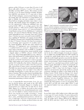
blood cells within 24 hours. A total of 231 cases were Figure 1
identified meeting MTP criteria. UK medical records Axial computed
(records of care at Queen Elizabeth Hospital Birming- tomography scan
ham [QEHB]) were present for 97 cases and, of these, demonstrating method
only 65 had undergone cross-sectional thoracic imag- of chest wall thickness
ing during their first admission to Camp Bastion hos- measurement (34mm).
pital or QEHB. One case was excluded as a result of
delay between imaging and injury (more than 1 year),
and the only female patient was excluded as this was
not felt representative following published data sug-
gesting female CWT is, on average, greater than that Figure 2 Axial computed tomography images demonstrating
of male patients. 9,10 This process yielded a final cohort the two methods of distance measurement to critical
structures at the left fifth intercostal space, midclavicular line.
of 63 patients. Most scans (n = 61) were performed in (A) Insertion perpendicular to skin (122mm). (B) Worst-case
Camp Bastion using a 64-slice Multidetector LightSpeed scenario angular insertion (60mm).
Volume CT (General Electric Medical Systems; http://
www3.gehealthcare.com) acquiring 5mm and 1.25mm (A)
slices. A further two scans were performed at QEHB us-
ing a LightSpeed Multislice CT scanner (General Elec-
16
tric Medical Systems) and a SOMATOM Definition AS
64-slice CT Scanner (Siemens AG; http://www.siemens.
com) following rapid evacuation of casualties back to
the United Kingdom. All but two scans (which were
pulmonary CT angiograms) were traumagrams using
a modified Baltimore protocol. Measurements were (B)
11
taken from postcontrast circle of Willis to symphysis
pubis soft-tissue reconstructions, where available, using predicted rate of injury to critical structures (Tables 1
a digital caliper on IMPAX software (AGFA Health- and 2) for five different lengths of needle for compari-
care Corp.; http://www.agfahealthcare.com). Measure- son. A failure was recorded when the CWT equaled or
ments were performed by a trainee military doctor after exceeded the needle length and an injury was recorded
instruction from a consultant radiologist who inde- when the needle length equaled or exceeded the distance
pendently verified all measurements to ensure reproduc- to a critical structure (shown for both perpendicular and
ibility. Axial images were used to locate the 2ICS MCL closest measurements). Needle lengths presented in Ta-
and the 5ICS midaxillary line (MAL), bilaterally, count- ble 1 range from a standard angiocatheter needle length
ing ribs, using the superior border of the third and sixth (45mm) to the first length at which measurements ex-
ribs, respectively, to ensure correct intercostal location. ceeded CWT (implying no failures). Needle lengths
The MAL was again located by marking the midpoint of presented in Table 2 range from the longest length at
the axilla on imaging. Care was made in slice selection which no measurements equaled the depth to a critical
and MAL identification to ensure the most correct ana- structures (implying no injury) to the length of the Air
tomical location. This was done despite knowledge that, Release System (North American Rescue, LLC; http://
in practice, in highly charged and often dangerous situ- www.narescue.com) currently used in UK military prac-
ations under which these devices are placed, textbook tice (80mm). Analysis was performed using GraphPad
anatomical positioning is seldom achieved. statistical software (GraphPad Software, Inc.; http://
www.graphpad.com). The Fisher exact test was used to
A set of perpendicular measurements, from skin sur- compare the total number of cases in each group (5ICS
face to parietal pleura, were made at the 2ICS MCL versus 2ICS and left side versus right) (Table 3) and
and 5ICS MAL, bilaterally (Figure 1). A further set of the unpaired Student t test was carried out to compare
measurements was made at the left-side 5ICS (L 5ICS) means.
MAL (Figure 2), as follows: the depth to first critical
structure if the device was inserted perpendicular to the Results
skin, and the depth to nearest critical structure if the
device was inserted at an angle, simulating a “worst- In our final cohort, all 63 subjects were white men with
case scenario.” Critical structures were defined as the a mean age of 24 years (median, 24 years; interquartile
heart, aorta, central/segmental pulmonary arterial ves- range [IQR], 21–27 years). Of these 63 casualties, there
sels, or diaphragm. Each set of measurements was then were 10 deaths (16%). Mean CWTs for the right-side
used, respectively, to tabulate predicted failure rate and 2ICS (R 2ICS) and then L 2ICS and R 5ICS and then
Optimal Device Length for Needle Thoracostomy 61