
Page 93 - Journal of Special Operations Medicine - Summer 2015
P. 93
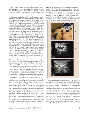
comfort. Widely prepare the skin over the anterior and right   Figure 3  Ultrasound-guided, right-sided stellate ganglion
              side of the neck with an alcohol and chlorhexidine solution,   block (SGB). (A) Transducer positioning during a long-axis
              and allow the solution to dry for 1 minute. Perform a clinical   (in-plane) approach to the stellate ganglion. (B) Path of the
              “time out” to confirm correct patient, correct procedure, and   needle, going through the sternocleidomastoid, under the
              correct side.                                      internal jugular vein, through the longus capitus muscle just
                                                                 ventral to the anterior tubercle of C6, and into the ventral
              4.  Ultrasound  positioning:  Apply a  small  amount,  approxi-  fascia of the longus coli muscle, laying immediately ventral to
              mately 2 grams, of sterile ultrasound gel to the anterior neck   the body of the C6 vertebra. (C) Long-axis needle approach,
              at the level of the cricoid membrane. While seated on the   with the needle tip in the ventral fascia of longus coli.
              right side of the patient, place a cleaned and prepared high-
              frequency linear transducer transverse at the level of the cricoid
              membrane (i.e., sixth cervical vertebra, or C6, level). Raising
              the procedure table to about the level of the provider’s chest
              usually facilitates proper ergonomics. The depth of the ultra-
              sound unit is set to visualize the ventral border of the C6 verte-
              bral body (usually 4 cm in male patients). Identify the anterior                           A
              tubercle of the C6 vertebra. The anterior tubercle of C6 has a
              distinct peaked appearance, and the level can be confirmed by
              being both at the level of the cricoid membrane and by it being
              the most caudad anterior tubercle (which can be confirmed
              with a short-axis slide in a caudad direction towards the clav-
              icle). Identify key landmarks: the common carotid artery, inte-
              rior jugular vein (facilitate viewing the entire internal jugular
              vein by having the patient perform the Valsalva maneuver),
              ventral portion of the C6 vertebral body, longus coli muscle
              overlying the vertebral body, longus capitus muscle (usually)
              overlying the anterior tubercle of C6. (Note: there is a high                              B
              degree of anatomic variation in the anterior neck.) While in
              this transverse view, use power Doppler or color Doppler to
              scan and identify vascular structures, especially looking for
              the well-documented anatomic variation of a vertebral artery
              coursing anterior and medial to the anterior tubercle of C6.

              5. Procedure: Refer to Figure 3. Envisioning a long-axis (or in-
              plane) lateral approach, mentally ensure the needle can reach
              the target area from the lateral neck. Place a skin wheal of
              0.5mL buffered 1% lidocaine at the needle entry site. Using a
              3.5-in long 22-gauge needle (or other appropriate needle), enter                           C
              the neck with the needle in long axis to the ultrasound trans-
              ducer (“in-plane” approach) going through sternocleidomas-
              toid, continuing just ventral to the tip of the anterior tubercle of
              C6, then continuing on until the needle tip has just penetrated
              the ventral fascia of longus coli, just medial to the longus capi-
              tus muscle and dorsal to the common carotid artery. The cervi-
              cal sympathetic chain usually courses along the ventral fascia
              of longus coli at this level, and it is sometimes, but not always,
              clearly visible on ultrasound. Initially aspirate to check for no   6.  Observation and monitoring: Observe and monitor the
              blood in the hub of the needle, then slowly inject 7–8mL 0.5%   patient for at least 30 minutes after completion of the injec-
              ropivacaine (over 2 minutes in 0.5mL aliquots) to mitigate risk   tion. Have the patient remain in the supine position (a pillow
              associated with potential intravascular injection. The (anechoic)   may be used at this time). The first sign of a successful block
              injectate should flow just dorsal to the ventral fascia of longus   will often be a sensation change on the right side of the face.
              coli. There is significant anatomic variation in the anterior neck   Once signs and symptoms of Horner’s syndrome are evident,
              and slight variations of this description may be required. Let the   the patient may sit reclined at a 20° angle for the remaining
              patient know that they can talk during the injection if needed.   observation period. These positions may facilitate produc-
              Periodically ask the patient during the injection if they are do-  tive anesthetic spread. Record the patient’s initial response to
              ing well, and let the patient know that questioning them is just   the injection, the time at which an obvious Horner’s response
              another way to monitor how they are doing. It is absolutely   was evident, and the quantitative score of the Horner’s syn-
              critical to constantly keep the needle tip in view. If the needle   drome. Approximately 20 minutes after the Horner’s response
              tip cannot be visualized, stop the injection, reacquire needle tip   is evident, inquire how the patient feels “mentally.” Usually
              visualization, aspirate while checking for no blood in the hub of   patients report some variation of feeling “relaxed, light, and
              the needle, and only then restart the injection.   calm.” An additional but optional step is to help the patient


              Guidelines for Stellate Ganglion Block for PTSD Anxiety                                         83