
Page 110 - Journal of Special Operations Medicine - Summer 2015
P. 110
plasma within 6 hours. Depending on when the test hours to determine the peak CK level and trend change
8
is performed, the potential for false measurements is over time.
significant and thus it is difficult to make clinical judg-
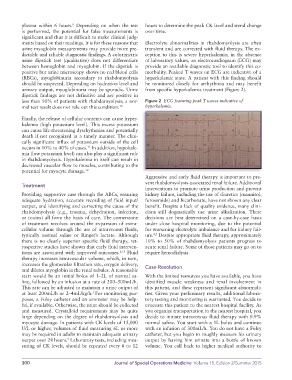
ments based on their readings. It is for these reasons that Electrolyte abnormalities in rhabdomyolysis are often
urine myoglobin measurements may provide more pre- transient and are corrected with fluid therapy. The ex-
dictable and reliable diagnostic findings. A colorimetric ception to this is severe hyperkalemia; in the absence
urine dipstick test (qualitative) does not differentiate of laboratory values, an electrocardiogram (ECG) may
between hemoglobin and myoglobin. If the dipstick is provide an available diagnostic tool to identify this co-
positive but urine microscopy shows no red blood cells morbidity. Peaked T waves on ECG are indicative of a
(RBCs), myoglobinuria secondary to rhabdomyolysis hyperkalemic state. A patient with this finding should
should be suspected. Depending on hydration level and be monitored closely for arrhythmia and may benefit
urinary output, myoglobinuria may be sporadic. Urine from specific hyperkalemia treatment (Figure 2).
dipstick findings are not definitive and are positive in
less than 50% of patients with rhabdomyolysis; a nor- Figure 2 ECG featuring peak T waves indicative of
mal test result does not rule out this condition. 10 hyperkalemia.
Finally, the release of cellular contents can cause hyper-
kalemia (high potassium level). This excess potassium
can cause life-threatening dysrhythmias and potentially
death if not recognized in a timely manner. The clini-
cally significant influx of potassium outside of the cell
occurs in 10% to 40% of cases. In addition, hypokale-
11
mia (low potassium level) can also play a significant role
in rhabdomyolysis. Hypokalemia in itself can result in
decreased vascular flow to muscles, contributing to the
potential for myocyte damage. 12
Aggressive and early fluid therapy is important to pre-
vent rhabdomyolysis-associated renal failure. Additional
Treatment
interventions to promote urine production and prevent
Providing supportive care through the ABCs, ensuring kidney failure, including the use of diuretics (mannitol,
adequate hydration, accurate recording of fluid input/ furosemide) and bicarbonate, have not shown any clear
output, and identifying and correcting the cause of the benefit. Despite a lack of quality evidence, many clini-
rhabdomyolysis (e.g., trauma, dehydration, infection, cians still dogmatically use urine alkalization. These
or toxins) all form the basis of care. The cornerstone decisions are best determined on a case-by-case basis
of treatment revolves around the expansion of extra- under close hospital monitoring, due to the potential
cellular volume through the use of intravenous fluids, for worsening electrolyte imbalance and for kidney fail-
typically normal saline or Ringer’s lactate. Although ure. Despite appropriate fluid therapy, approximately
4,5
there is no clearly superior specific fluid therapy, ret- 10% to 50% of rhabdomyolysis patients progress to
rospective studies have shown that early fluid interven- acute renal failure. Some of those patients may go on to
tions are associated with improved outcomes. 5,13 Fluid require hemodialysis.
therapy increases intravascular volume, which, in turn,
increases the glomerular filtration rate, oxygen delivery, Case Resolution
and dilutes myoglobin in the renal tubules. A reasonable
start would be an initial bolus of 1–2L of normal sa- With the limited resources you have available, you have
line, followed by an infusion at a rate of 200–500mL/h. identified muscle weakness and renal involvement in
This rate can be adjusted to maintain a urine output of this patient, and these represent significant abnormali-
at least 200mL/h or 2–4mL/kg/h. For monitoring pur- ties. Given your preliminary results, additional labora-
5
poses, a Foley catheter and an urometer may be help- tory testing and monitoring is warranted. You decide to
ful, if available. Otherwise, the urine should be collected evacuate this patient to the nearest hospital facility. As
and measured. Crystalloid requirements may be quite you organize transportation to the nearest hospital, you
large depending on the degree of rhabdomyolysis and decide to initiate intravenous fluid therapy with 0.9%
myocyte damage. In patients with CK levels of 15,000 normal saline. You start with a 1L bolus and continue
U/L or higher, volumes of fluid measuring 6L or more with an infusion of 300mL/h. You do not have a Foley
may be required in adults to maintain adequate urinary catheter, but you begin to roughly measure his urinary
output over 24 hours. Laboratory tests, including mea- output by having him urinate into a bottle of known
5
suring of CK levels, should be repeated every 6 to 12 volume. You call back to higher medical authority to
100 Journal of Special Operations Medicine Volume 15, Edition 2/Summer 2015