
Page 124 - Journal of Special Operations Medicine - Spring 2015
P. 124
associated with this complaint. He denied any changes in • Comedo: retained secretions of material within the
washing detergents, soaps, or use of lotions, and denied pilosebaceous follicle.
any known exposure to plants or animals in the area • Pustule: a circumscribed elevation containing pus.
prior to presentation. Physical examination was unre • Cyst: a circumscribed, thick walled, slightly elevated
markable with the exception of the skin findings on the lesion extending into the deep dermis and subcutane
posterior aspect of his left knee. The patient was able to ous fat.
bear weight without difficulty, had a normal knee joint • Wheal or hive: a distinctive white to pink or pale red,
examination, and no lymphadenopathy noted. He was edematous, solid elevation formed by local, superfi
afebrile with normal vital signs. cial, transient edema. They characteristically disap
pear, yet may reappear within a period of hours.
• Telangiectasia: blanchable (fades with fingertip pres
Review of the Dermatologic Evaluation
sure), small, superficial, dilated capillaries.
It has been said that the eye cannot see what the mind does • Purpura: nonblanchable, purple area of the skin that
not know. It is difficult to accurately describe a dermato may be flat or raised.
logic abnormality without having a basic knowledge of
terminology. The following is a review of dermatologic Secondary lesions
terminology and lesion types (Figure 2) from the Special Description of secondary lesions does not offer the same
Operations Forces Medical Handbook. 1 diagnostic descriptive power as the primary lesions, but
represents the evolution of the primary lesions.
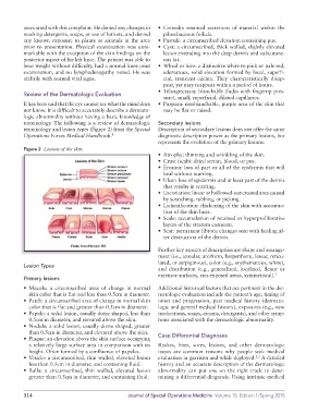
Figure 2 Lesions of the skin.
• Atrophy: thinning and wrinkling of the skin.
• Crust (scab): dried serum, blood, or pus.
• Erosion: loss of part or all of the epidermis that will
heal without scarring.
• Ulcer: loss of epidermis and at least part of the dermis
that results in scarring.
• Excoriation: linear or hollowedout crusted area caused
by scratching, rubbing, or picking.
• Lichenification: thickening of the skin with accentua
tion of the skin lines.
• Scale: accumulation of retained or hyperproliferative
layers of the stratum corneum.
• Scar: permanent fibrotic changes seen with healing af
ter destruction of the dermis.
Further key aspects of description are shape and arrange
ment (i.e., annular, arciform, herpetiform, linear, reticu
lated, or serpiginous), color (e.g., erythematous, white),
Lesion Types
and distribution (e.g., generalized, localized, flexor or
extensor surfaces, sunexposed areas, symmetrical). 1
Primary lesions
• Macule: a circumscribed area of change in normal Additional historical factors that are pertinent in the der
skin color that is flat and less than 0.5cm in diameter. matologic evaluation include the patient’s age, timing of
• Patch: a circumscribed area of change in normal skin onset and progression, past medical history (dermato
color that is flat and greater than 0.5cm in diameter. logic and general medical history), exposures (e.g., new
• Papule: a solid lesion, usually dome shaped, less than medications, soaps, creams, detergents), and other symp
0.5cm in diameter, and elevated above the skin. toms associated with the dermatologic abnormality.
• Nodule: a solid lesion, usually dome shaped, greater
than 0.5cm in diameter, and elevated above the skin.
• Plaque: an elevation above the skin surface occupying Case Differential Diagnoses
a relatively large surface area in comparison with its Rashes, bites, sores, lesions, and other dermatologic
height. Often formed by a confluence of papules. issues are common reasons why people seek medical
• Vesicle: a circumscribed, thin walled, elevated lesion evaluation in garrison and while deployed. A detailed
2,3
less than 0.5cm in diameter, and containing fluid. history and an accurate description of the dermatologic
• Bulla: a circumscribed, thin walled, elevated lesion abnormality can put one on the right track to deter
greater than 0.5cm in diameter, and containing fluid. mining a differential diagnosis. Using intrinsic medical
114 Journal of Special Operations Medicine Volume 15, Edition 1/Spring 2015