
Page 32 - Journal of Special Operations Medicine - Summer 2014
P. 32
platform. The SAM Pelvic Sling previously had been control. Hemorrhage control was defined as a minimum
®
developed in collaboration with Legacy Health Systems of 45 seconds of continuous blood flow cessation.
under military funding (Office of Naval Research Grant
N00014-01-1-0132) and is a pelvic binder indicated for The experimental control was manual pressure alone at
the reduction of displaced, open-book–type pelvic frac- the target point to occlude the artery. The pressure was
tures. Cadaveric and human clinical studies have dem- recorded at the time of hemorrhage control.
onstrated that there is a specific range of force that is
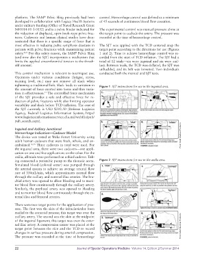
most effective in reducing pubic symphysis diastasis in The SJT was applied with the TCD centered atop the
patients with pelvic fractures while maintaining patient target point according to the directions for use (Figures
safety. 9,10 For this safety reason, the SAM Pelvic Sling 1 and 2). Time to achieve hemorrhage control was re-
®
(and now also the SJT) incorporates a mechanism that corded from the start of TCD inflation. The SJT had a
limits the applied circumferential tension to the thresh- total of 12 trials—six were inguinal and six were axil-
old amount. lary. Between trials, the TCD was deflated, the SJT was
unbuckled, and its belt was loosened. Two individuals
This control mechanism is relevant to tourniquet use. conducted both the manual and SJT tests.
Operators under various conditions (fatigue, stress,
training level, etc.) may not remove all slack when
tightening a traditional belt. Slack leads to variation in Figure 1 SJT instructions for use in the inguinal area.
the amount of force exerted into tissue and thus varia-
tion in effectiveness. The controlled force mechanism
11
of the SJT provides a safe and effective force for re-
duction of pelvic fractures while also limiting operator
variability and slack before TCD inflation. The cost of
the SJT currently is USD $292.50 (Defense Logistics
Agency, Federal Logistics Information System; http://
www.logisticsinformationservice.dla.mil/webflis/pub/
pub_search.aspx).
Inguinal and Axillary Junctional
Hemorrhage Indication—Cadaver Model
The device was tested at Wake Forest University using From SAM Medical Products
adult human cadavers that were fresh, whole, and un-
embalmed. 12,13 Three cadavers in total were used. For
the inguinal area, there were two cadavers—one appli-
cation on one and five applications on the other. For the
axilla, all trials were performed on a third cadaver. Tub-
ing connected a peristaltic pump to the thoracic aorta. Figure 2 SJT instructions for use in the axilla area.
Simulated blood (colored water) was pumped through
the arterial system to achieve an average arterial flow
rate of 330mL/min, which approximates normal flow
through the axillary and external iliac arteries. The bra-
chial artery was opened to allow bleeding and to moni-
tor blood flow continuously through the axillary artery.
Similarly, the popliteal artery was opened to bleeding
and to monitor blood flow continuously through the ex-
ternal iliac and femoral arteries.
There were two target points for the application of pres-
sure. The first was the skin of the infraclavicular fossa
medial to the coracoid process; this target was over the
axillary artery. The second was the skin at the midpoint
of the inguinal ligament; this target was over the exter-
nal iliac artery. A compression sensor was placed at the From SAM Medical Products
target point between the skin and the TCD to record
changes in surface pressure during arterial compression.
The pressure was recorded at the time of hemorrhage
22 Journal of Special Operations Medicine Volume 14, Edition 2/Summer 2014