
Page 17 - Journal of Special Operations Medicine - Summer 2014
P. 17
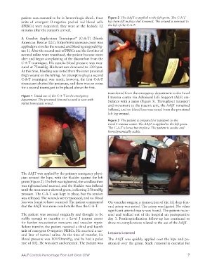
patient was assessed to be in hemorrhagic shock. Four   Figure 2  The AAJT is applied to the left groin. The C-A-T
              units of emergent O-negative packed red blood cells   has been left in place but loosened. The wound is seen just to
              (PRBCs) were requested; they were at the bedside 12   the left of the C-A-T.
              minutes after the patient’s arrival.

              A Combat Application Tourniquet  (C-A-T) (North
                                             ®
              Ameri  can Rescue LLC; http://www.narescue.com) was
                applied proximal to the wound, and bleeding stopped (Fig-
              ure 1). After the second unit of PRBCs and the first liter of
              normal saline were transfused, the patient became more
              alert and began complaining of the discomfort from the
              C-A-T tourniquet. His systolic blood pressure was mea-
              sured at 75mmHg. His heart rate decreased to 130 bpm.
              At this time, bleeding was noted from the inner proximal
              thigh wound on the left leg. An attempt to place a second
              C-A-T tourniquet was made; however, the first C-A-T
              tourniquet abutted the perineum, and there was no room
              for a second tourniquet to be placed above the first.
                                                                 transferred from the emergency department to the Level
              Figure 1  Initial use of the C-A-T in the emergency   I trauma center via Advanced Life Support (ALS) am-
              department. The proximal femoral wound is seen with    bulance with a nurse (Figure 3). Throughout transport
              initial hemostasis noted.
                                                                 and movement to the trauma unit, the AAJT remained
                                                                 inflated, and no blood loss was noted from the proximal
                                                                 left leg wound.
                                                                 Figure 3  The patient is prepared for transport to the
                                                                 Level 1 trauma center. The AAJT is applied to the left groin.
                                                                 The C-A-T is loose but in place. The patient is awake and
                                                                 hemodynamically stable.


              The AAJT was applied by the primary emergency physi-
              cian around the hips, with the bladder against the left
              groin (Figure 2). The belt was tightened, the windlass then
              was tightened and secured, and the bladder was inflated
              until the manometer showed green, indicating 250mmHg
              pressure. The C-A-T was kept in place, but the tension
              was released. The wounds were reassessed, and no blood
              loss was found to have occurred. The patient commented   On vascular surgery, a transsection of the left deep fem-
              that the AAJT was more comfortable than the C-A-T.  oral artery was noted. The artery was ligated. No other
                                                                 significant arterial injury was found. The patient recov-
              The patient was assessed surgically and thought to be   ered and walked out of the hospital on postoperative
              stable enough to transfer to a Level I trauma center   day 3. Posthospitalization follow-up has continued to
              for further resuscitative measures and vascular repair.   show no complications related to the use of the AAJT.
              Before transfer, the patient received a third and fourth
              unit of emergent O-negative PRBCs. He received a sec-
              ond liter of normal saline. At the time of transfer, his   Lessons Learned
              blood pressure was 101/50mmHg, and he had a pulse   The AAJT was quickly applied over the hips and po-
              rate of 102. He was alert and oriented. The patient was   sitioned over the groin. Slack removal is essential for


              AAJT Controls Hemorrhage From Left Groin GSW                                                     7