
Page 98 - ATP-P 11th Ed
P. 98
e. If SVPs are initially present and can no longer be seen on subsequent examinations, the
provider should be concerned for increasing ICP.
SECTION 1 Technique
1. Gently lift the eyelid until the pupil is in view.
2. Using a handheld ophthalmoscope, the provider should maneuver himself or herself to
a position where the optic disc can be visualized.
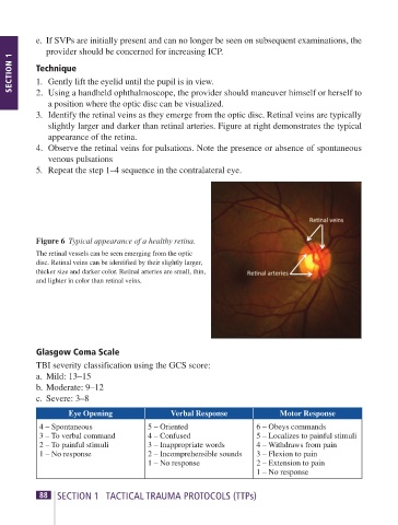
3. Identify the retinal veins as they emerge from the optic disc. Retinal veins are typically
slightly larger and darker than retinal arteries. Figure at right demonstrates the typical
appearance of the retina.
4. Observe the retinal veins for pulsations. Note the presence or absence of spontaneous
venous pulsations
5. Repeat the step 1–4 sequence in the contralateral eye.
Figure 6 Typical appearance of a healthy retina.
The retinal vessels can be seen emerging from the optic
disc. Retinal veins can be identified by their slightly larger,
thicker size and darker color. Retinal arteries are small, thin,
and lighter in color than retinal veins.
Glasgow Coma Scale
TBI severity classification using the GCS score:
a. Mild: 13–15
b. Moderate: 9–12
c. Severe: 3–8
Eye Opening Verbal Response Motor Response
4 – Spontaneous 5 – Oriented 6 – Obeys commands
3 – To verbal command 4 – Confused 5 – Localizes to painful stimuli
2 – To painful stimuli 3 – Inappropriate words 4 – Withdraws from pain
1 – No response 2 – Incomprehensible sounds 3 – Flexion to pain
1 – No response 2 – Extension to pain
1 – No response
88 SECTION 1 TACTICAL TRAUMA PROTOCOLS (TTPs) ATP-P Handbook 11th Edition 89