
Page 97 - ATP-P 11th Ed
P. 97
10. Repeat the previous sequence in the opposite eye. Annotate both ONSDs on the PFC
Casualty Card.
11. ONSDs should be obtained, when possible, at regular intervals to help assess changes
in ICP, particularly when the neurologic examination is poor and/or unreliable (i.e., SECTION 1
with sedation). Serial measurements with progressive diameter enlargement and/
or asymmetry in ONSDs should be considered indicative of worsening intracranial
hypertension.
CAUTION: ONSD measurements are contraindicated in eye injuries. NEVER apply pres-
sure to an injured eye.
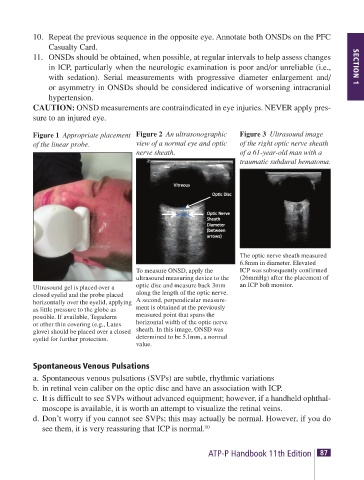
Figure 1 Appropriate placement Figure 2 An ultrasonographic Figure 3 Ultrasound image
of the linear probe. view of a normal eye and optic of the right optic nerve sheath
nerve sheath. of a 61-year-old man with a
traumatic subdural hematoma.
The optic nerve sheath measured
6.8mm in diameter. Elevated
To measure ONSD, apply the ICP was subsequently confirmed
ultrasound measuring device to the (26mmHg) after the placement of
Ultrasound gel is placed over a optic disc and measure back 3mm an ICP bolt monitor.
closed eyelid and the probe placed along the length of the optic nerve.
horizontally over the eyelid, applying A second, perpendicular measure-
as little pressure to the globe as ment is obtained at the previously
possible. If available, Tegaderm measured point that spans the
or other thin covering (e.g., Latex horizontal width of the optic nerve
glove) should be placed over a closed sheath. In this image, ONSD was
eyelid for further protection. determined to be 5.1mm, a normal
value.
Spontaneous Venous Pulsations
a. Spontaneous venous pulsations (SVPs) are subtle, rhythmic variations
b. in retinal vein caliber on the optic disc and have an association with ICP.
c. It is difficult to see SVPs without advanced equipment; however, if a handheld ophthal-
moscope is available, it is worth an attempt to visualize the retinal veins.
d. Don’t worry if you cannot see SVPs; this may actually be normal. However, if you do
see them, it is very reassuring that ICP is normal. 10
86 SECTION 1 TACTICAL TRAUMA PROTOCOLS (TTPs) ATP-P Handbook 11th Edition 87