
Page 30 - Journal of Special Operations Medicine - Fall 2016
P. 30
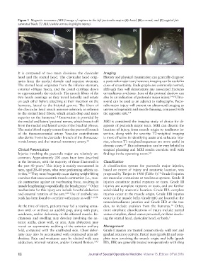
Figure 1  Magnetic resonance (MRI) image of rupture in the left pectoralis major. (A) Axial, (B) coronal, and (C) sagittal fat-
            saturated blade T2 MRI (white arrows highlight injury).

                                         (A)                                    (B)                        (C)


            It is composed of two main divisions: the clavicular   Imaging
            head and the sternal head. The clavicular head origi-  History and physical examination can generally diagnose
            nates  from  the  medial  clavicle  and  superior  sternum.   a pectoralis major tear; however, imaging can be useful in
            The sternal head originates from the inferior sternum,   cases of uncertainty. Radiographs are commonly normal,
            external oblique fascia, and the costal cartilage down   although they will demonstrate any associated fractures
            to approximately the sixth rib. The muscle fibers of the   or tendinous avulsions. Loss of the pectoral shadow can
            two heads converge as they travel laterally and rotate   also be an indication of pectoralis major injury. 2–4,6  Ultra-
            on each other before attaching at their insertion on the   sound can be used as an adjunct to radiographs. Pecto-
            humerus, lateral to the bicipital groove. The fibers of   ralis major injury will present on ultrasound imaging as
            the clavicular head attach anterior-inferiorly in relation   uneven echogenicity and muscle thinning, compared with
            to the sternal head fibers, which attach deep and more   the opposite side. 2–4
            superior on the humerus.  Innervation is provided by
                                  1–4
            the medial and lateral pectoral nerves, which branch off   MRI is considered the imaging study of choice for di-
            from the medial and lateral cords of the brachial plexus.   agnosis of pectorals major tears. MRI can discern the
            The main blood supply comes from the pectoral branch   location of injury, from muscle origin to tendinous in-
            of the thoracoacromial  artery. Vascular  contributions   sertion, along with the severity. T2-weighted imaging
            also derive from the clavicular branch of the thoracoac-  is most effective in identifying acute and subacute inju-
            romial artery and the internal mammary artery. 3,4  ries, whereas T1-weighted sequences are more useful in
                                                               chronic cases.  This information can be very helpful in
                                                                           3,4
            Clinical Presentation                              surgical planning and MRI results correlate well with
            Injuries  involving  the  pectoralis  major  are  relatively  un-  findings in the operating room. 3,8
            common. Approximately 200 cases have been described
            in the literature, with the majority of those illustrated in   Classification
            the past 40 years.  This injury is mainly encountered in   A classification system for pectoralis major injuries,
                           5
            men, aged 20–40 years, who were performing athletic ac-  based on extent of injury and anatomic location, was
            tivities.  They most frequently occur during weight-lifting   proposed by Tietjen in 1980 (Table 1).  Grade I injuries
                  3,4
                                                                                                12
            exercises that cause eccentric muscle contraction (i.e., mus-  are muscular contusions or tendinous sprains. Grade II
            cle contraction against an overbearing force, resulting in   injuries  constitute  partial  ruptures  or  tears.  Grade  III
            muscle lengthening)—specifically, the bench press.  Other   injuries are complete ruptures or tears, and are further
                                                     1–9
            mechanisms for this injury can include forceful abduction   subdivided by anatomic location. Grade IIIA complete
            and external rotation of the arm. 2,10  Use of anabolic ste-  injuries occur at the muscle origin. Grade IIIB injuries
            roids has been found to correlate with injury as well. 2–6,9,11  occur in the muscle belly. Grade IIIC are located at the
                                                               musculotendinous junction and Grade IIID at the ten-
            At the time of injury, patients may feel a tearing sensa-  don, to include avulsion from the humerus.  Other,
                                                                                                       12
            tion with or without an audible pop, along with pain,   more simplistic classifications of injury include partial
            weakness, and/or deformity of the affected muscle. Ec-  versus complete, distal versus proximal, or those involv-
            chymosis and swelling may develop involving the an-  ing the sternal head, clavicular head, or both. 3
            terior  axilla,  chest  wall,  or  arm.  Arm  abduction  may
            reveal an asymmetric webbing of the anterior axillary   Management
            fold, compared with the unaffected  side. Chest defor-  Grade I injuries are treated conservatively with rest and
            mity may also be accentuated with contracted arm ad-  gradual return to activity. Partial tears (grade II) and com-
            duction. Pain and weakness may be elicited with arm   plete tears involving the muscle origin and belly (grade
            adduction, internal rotation, and/or forward flexion. 3,4,7  IIIA, IIIB) are generally treated nonoperatively with sling


            12                                        Journal of Special Operations Medicine  Volume 15, Edition 3/Fall 2016