
Page 130 - Journal of Special Operations Medicine - Spring 2015
P. 130
differentiating between gynecologic pathologies can be
VISCERAL PAIN very difficult.
Visceral nerve fibers are primitive nerve tissue that
innervates hollow organs. When stimulated, the Classic physical findings all are variable in sensitivity
pain felt by visceral nerves is often vague and mid and specificity but include:
line or poorly localized. The pain is often difficult
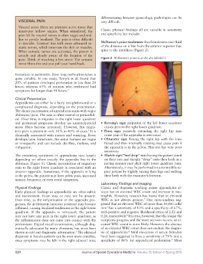
to describe. Contrast this with more advanced so McBurney’s point tenderness: focal tenderness onethird
matic nerves, which innervate the skin or muscles. of the distance on a line from the anterior superior iliac
When somatic nerves are activated, the person is spine to the umbilicus (Figure 2).
acutely and clearly aware of the location of the
pain. Think of touching a hot stove: The somatic Figure 2 McBurney’s point is at the dot labeled 1.
nerve fibers fire and you pull your hand back.
formation or peritonitis. How long perforation takes is
quite variable. In one study, Temple et al. found that
20% of patients developed perforation in less than 24
hours, whereas 65% of patients who perforated had 2
symptoms for longer than 48 hours. 4
Clinical Presentation
Appendicitis can either be a fairly straightforward or a 1
complicated diagnosis, depending on the presentation.
The classic presentation of appendicitis starts with vague 3
abdominal pain. The pain is often central or periumbili
cal. Over time, it migrates to the right lower quadrant
and peritoneal symptoms develop (as somatic/parietal • Rovsing’s sign: palpation of the left lower quadrant
nerve fibers become activated). However, this migra causes pain in the right lower quadrant.
tory pain is present in only 50% to 60% of cases. It is • Psoas sign: passively extending the right hip may
5
classically associated with nausea and vomiting. Fever cause pain if the appendix is retrocecal.
develops later. Sometimes, the initial features are vague • Obturator sign: flexing the right hip with the knee
or nonspecific and can include diarrhea, malaise, and flexed and then internally rotating may cause pain if
indigestion. the appendix is in the pelvis. This test has very poor
sensitivity.
The presenting symptoms of appendicitis vary largely • Markle sign/“heel drop” test: having the patient stand
depending on where exactly the appendix lies in the on their toes and sharply “drop” onto their heels in a
abdomen (Figure 1). Classic presentation of migratory jarring manner may elicit right lower quadrant pain.
pain to the right lower quadrant is associated with an Alternatively, it may be performed on a nonmobile su
anterior appendix. Sometimes, if the appendix is lying pine patient by slightly raising their legs and striking
in the pelvis, the patient may have pelvic pain, increased their heels with the examiner’s forearm.
urinary frequency, or even rectal symptoms.
Laboratory Findings and Imaging
Physical Findings Classic and dogmatic teaching insists appendicitis al
Early physical findings in appendicitis are often subtle ways has an elevated WBC count and increase in neu
and inconsistent. Fever may or may not be present. trophils. However, research has found that an elevated
7
Over time, as the inflammation of the appendix pro WBC is not always present. One metaanalysis sug
gresses, the peritoneum (anterior position) may become gested that an elevated WBC of more than 10,000 cells/
3
inflamed, causing localized tenderness in the right lower mm has a sensitivity of 83% and a specificity of 67%,
quadrant. If the appendix is retrocecal, the patient with positive and negative likelihood ratios of 2.52 and
may not have any pain in the right lower quadrant, as 0.26, respectively. It is true, however, that the longer the
8
the inflammation does not come into contact with the symptoms progress and the more necrosis occurs, an el
peritoneum. Digital rectal examination, although dog evated WBC count is more likely. However, the absence
matically advocated by many clinicians, has never been of an elevated WBC count does not exclude the diagno
8
shown to add any diagnostic information. The physical sis of appendicitis. Mild elevations in serum bilirubin
6
diagnosis in female patients can be even more complex, have been suggested to have a sensitivity of 70% and a
9
since symptoms may be felt in the right adnexal area; specificity of 86% for appendiceal perforation. Most
120 Journal of Special Operations Medicine Volume 15, Edition 1/Spring 2015