
Page 12 - Journal of Special Operations Medicine - Spring 2015
P. 12
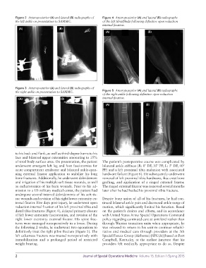
Figure 2 Anteroposterior (A) and lateral (B) radiographs of Figure 4 Anteroposterior (A) and lateral (B) radiographs
the left ankle on presentation to SAMMC. of the left tibia/fibula following definitive open reduction
internal fixation.
(A) (B)
(A) (B)
Figure 3 Anteroposterior (A) and lateral (B) radiographs of
the right ankle on presentation to SAMMC. Figure 5 Anteroposterior (A) and lateral (B) radiographs
of the right ankle following definitive open reduction
(A) (B) internal fixation.
(A) (B)
to his back and flank, as well as thirddegree burns to his
face and bilateral upper extremities amounting to 15%
of total body surface area. On presentation, the patient The patient’s postoperative course was complicated by
underwent emergent left leg and foot fasciotomies for bilateral ankle stiffness (R: 0º DF, 35º PF; L: 5º DF, 40º
acute compartment syndrome and bilateral anklespan PF) and a left proximal tibia malunion with associated
ning external fixator application to stabilize his long hardware failure (Figure 6). He subsequently underwent
bone fractures. Additionally, he underwent debridement removal of left proximal tibia hardware, iliac crest bone
and irrigation of his multiple soft tissue wounds, as well grafting, and application of a ringed external fixator.
as escharotomies of his burn wounds. Prior to his ad The ringed external fixator was removed several months
mission to a US military medical center, the patient had later after he had healed his proximal tibia fracture.
undergone several interval debridements of his soft tis
sue wounds and revision of his right lower extremity ex Despite bony union of all of his fractures, he had con
ternal fixator. Five days postinjury, he underwent open tinued bilateral ankle pain and decreased ankle range of
reduction internal fixation of his left proximal tibia and motion, which significantly limited his function. Based
distal tibia fractures (Figure 4), delayed primary closure on the patient’s desires and efforts, and in accordance
of left lower extremity fasciotomies, and revision of the with United States Army Special Operations Command
right lower extremity external fixator. His spine frac policy regarding continued care at unit level rather than
tures were managed nonoperatively in a brace. During through Warrior transition units when appropriate, he
the following 2 weeks, he underwent two operations to was released to return to his unit to continue rehabil
definitively treat the right pilon fracture (Figure 5). The itative and medical care through providers at the 5th
left calcaneus fracture was treated nonoperatively with Special Forces Group (Airborne) [SFG(A)] based at Fort
immobilization and a prolonged period of restricted Campbell, Kentucky, at the earliest juncture that his
weight bearing. providers felt medically appropriate to do so. Despite
2 Journal of Special Operations Medicine Volume 15, Edition 1/Spring 2015